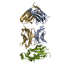
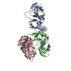
- PDB-4l6r: Structure of the class B human glucagon G protein coupled receptor -
+
Open data
ID or keywords:
Loading...
-
Basic information
Entry
Database: PDB / ID: 4l6r
Title
Structure of the class B human glucagon G protein coupled receptor
Components
Soluble cytochrome b562 and Glucagon receptor chimera
Keywords
MEMBRANE PROTEIN / Human glucagon receptor / diabetes / GPCR network / PSI-Biology / novel protein engineering / Structural Genomics / Protein Structure Initiative / GPCR / membrane
Function / homology
Function and homology information
regulation of glycogen metabolic process / glucagon receptor activity / response to starvation / peptide hormone binding / response to nutrient / cellular response to glucagon stimulus / guanyl-nucleotide exchange factor activity / cellular response to starvation / generation of precursor metabolites and energy / electron transport chain ...regulation of glycogen metabolic process / glucagon receptor activity / response to starvation / peptide hormone binding / response to nutrient / cellular response to glucagon stimulus / guanyl-nucleotide exchange factor activity / cellular response to starvation / generation of precursor metabolites and energy / electron transport chain / regulation of blood pressure / adenylate cyclase-modulating G protein-coupled receptor signaling pathway / Glucagon signaling in metabolic regulation / Glucagon-type ligand receptors / glucose homeostasis / adenylate cyclase-activating G protein-coupled receptor signaling pathway / G alpha (s) signalling events / G alpha (q) signalling events / periplasmic space / electron transfer activity / cell surface receptor signaling pathway / iron ion binding / heme binding / positive regulation of gene expression / membrane / plasma membrane Similarity search - Function
GPCR, family 2, glucagon receptor / GPCR, family 2, glucagon-like peptide-1/glucagon receptor / Cytochrome c/b562 / G-protein coupled receptors family 2 signature 1. / : / GPCR, family 2, extracellular hormone receptor domain / G-protein coupled receptors family 2 profile 1. / Domain present in hormone receptors / Hormone receptor domain / GPCR family 2, extracellular hormone receptor domain superfamily ...GPCR, family 2, glucagon receptor / GPCR, family 2, glucagon-like peptide-1/glucagon receptor / Cytochrome c/b562 / G-protein coupled receptors family 2 signature 1. / : / GPCR, family 2, extracellular hormone receptor domain / G-protein coupled receptors family 2 profile 1. / Domain present in hormone receptors / Hormone receptor domain / GPCR family 2, extracellular hormone receptor domain superfamily / G-protein coupled receptors family 2 signature 2. / GPCR, family 2, secretin-like, conserved site / GPCR, family 2, secretin-like / 7 transmembrane receptor (Secretin family) / Rhopdopsin 7-helix transmembrane proteins / Rhodopsin 7-helix transmembrane proteins / GPCR, family 2-like / G-protein coupled receptors family 2 profile 2. / Cytochrome b562 / Cytochrome b562 / Cytochrome c/b562 / Four Helix Bundle (Hemerythrin (Met), subunit A) / Up-down Bundle / Mainly Alpha Similarity search - Domain/homology
Resolution: 3.3→33.12 Å / Cor.coef. Fo:Fc: 0.8834 / Cor.coef. Fo:Fc free: 0.8424 / Cross valid method: THROUGHOUT / σ(F): 0 / Stereochemistry target values: Engh & Huber Details: 1. THE DIFFRACTION DATA ARE ANISOTROPIC. THE RESOLUTION LIMITS OF A*, B*, AND C* AXES ARE 3.3, 3.4 AND 3.3A, RESPECTIVELY. DIFFRAC DATA WERE INCLUDED IN REFINEMENT TO 3.3A IN THE A* AND C* ...Details: 1. THE DIFFRACTION DATA ARE ANISOTROPIC. THE RESOLUTION LIMITS OF A*, B*, AND C* AXES ARE 3.3, 3.4 AND 3.3A, RESPECTIVELY. DIFFRAC DATA WERE INCLUDED IN REFINEMENT TO 3.3A IN THE A* AND C* DIRECTIONS, WITH AN OVERALL EFFECTIVE AND REPORTED RESOLUTION OF 3.4A. 2. THE DENSITIES AT THE BOTTOM OF HELIX VI AND VII NEAR LYS349 WERE MODELLED AS A PEG-400 FRAGMENT (PEG) MOLECULE FROM THE CRYSTALLIZATION CONDITION.
Rfactor
Num. reflection
% reflection
Selection details
Rfree
0.3385
430
4.79 %
RANDOM
Rwork
0.2835
-
-
-
obs
0.2862
8981
91.59 %
-
Displacement parameters
Biso mean: 126.83 Å2
Baniso -1
Baniso -2
Baniso -3
1-
-1.1296 Å2
0 Å2
0 Å2
2-
-
21.5553 Å2
0 Å2
3-
-
-
-20.4256 Å2
Refine analyze
Luzzati coordinate error obs: 1.234 Å
Refinement step
Cycle: LAST / Resolution: 3.3→33.12 Å
Protein
Nucleic acid
Ligand
Solvent
Total
Num. atoms
3049
0
7
0
3056
Refine LS restraints
Refine-ID
Type
Dev ideal
Number
Restraint function
Weight
X-RAY DIFFRACTION
t_bond_d
0.01
3118
HARMONIC
2
X-RAY DIFFRACTION
t_angle_deg
1.04
4228
HARMONIC
2.5
X-RAY DIFFRACTION
t_dihedral_angle_d
1432
SINUSOIDAL
15
X-RAY DIFFRACTION
t_incorr_chiral_ct
X-RAY DIFFRACTION
t_pseud_angle
X-RAY DIFFRACTION
t_trig_c_planes
65
HARMONIC
2
X-RAY DIFFRACTION
t_gen_planes
459
HARMONIC
5
X-RAY DIFFRACTION
t_it
3118
HARMONIC
20
X-RAY DIFFRACTION
t_nbd
X-RAY DIFFRACTION
t_omega_torsion
2.79
X-RAY DIFFRACTION
t_other_torsion
1.81
X-RAY DIFFRACTION
t_improper_torsion
X-RAY DIFFRACTION
t_chiral_improper_torsion
403
SEMIHARMONIC
5
X-RAY DIFFRACTION
t_sum_occupancies
X-RAY DIFFRACTION
t_utility_distance
X-RAY DIFFRACTION
t_utility_angle
X-RAY DIFFRACTION
t_utility_torsion
X-RAY DIFFRACTION
t_ideal_dist_contact
3725
SEMIHARMONIC
4
LS refinement shell
Resolution: 3.3→3.69 Å / Total num. of bins used: 5
Rfactor
Num. reflection
% reflection
Rfree
0.3136
118
5.11 %
Rwork
0.2812
2190
-
all
0.283
2308
-
obs
-
2190
91.59 %
Refinement TLS params.
Method: refined / Refine-ID: X-RAY DIFFRACTION
ID
L11 (°2)
L12 (°2)
L13 (°2)
L22 (°2)
L23 (°2)
L33 (°2)
S11 (Å °)
S12 (Å °)
S13 (Å °)
S21 (Å °)
S22 (Å °)
S23 (Å °)
S31 (Å °)
S32 (Å °)
S33 (Å °)
T11 (Å2)
T12 (Å2)
T13 (Å2)
T22 (Å2)
T23 (Å2)
T33 (Å2)
Origin x (Å)
Origin y (Å)
Origin z (Å)
1
16.6309
2.3438
3.4299
6.0851
0.6799
14.8771
0.1704
-0.1676
-0.54
0.7744
-0.1591
-0.9671
-0.0046
0.2207
-0.0112
0.1043
-0.2881
-0.1636
0.047
0.1298
-0.5395
16.4176
4.5252
-2.0054
2
0.9466
0.6168
1.8062
1.2789
2.035
5.4002
0.3433
0.0555
-0.2635
0.1079
0.3087
-0.299
-0.3608
1.0885
-0.652
-0.379
-0.2048
0.0527
0.6079
-0.1883
-0.6079
16.1524
-11.4685
-44.8801
Refinement TLS group
ID
Refine-ID
Refine TLS-ID
Selection details
Auth asym-ID
Auth seq-ID
1
X-RAY DIFFRACTION
1
{ A|1001 - A|1106 }
A
1001 - 1106
2
X-RAY DIFFRACTION
2
{ A|123 - A|429 }
A
123 - 429
+
About Yorodumi
-
News
-
Feb 9, 2022. New format data for meta-information of EMDB entries
New format data for meta-information of EMDB entries
Version 3 of the EMDB header file is now the official format.
The previous official version 1.9 will be removed from the archive.
In the structure databanks used in Yorodumi, some data are registered as the other names, "COVID-19 virus" and "2019-nCoV". Here are the details of the virus and the list of structure data.
Jan 31, 2019. EMDB accession codes are about to change! (news from PDBe EMDB page)
EMDB accession codes are about to change! (news from PDBe EMDB page)
The allocation of 4 digits for EMDB accession codes will soon come to an end. Whilst these codes will remain in use, new EMDB accession codes will include an additional digit and will expand incrementally as the available range of codes is exhausted. The current 4-digit format prefixed with “EMD-” (i.e. EMD-XXXX) will advance to a 5-digit format (i.e. EMD-XXXXX), and so on. It is currently estimated that the 4-digit codes will be depleted around Spring 2019, at which point the 5-digit format will come into force.
The EM Navigator/Yorodumi systems omit the EMD- prefix.
Related info.:Q: What is EMD? / ID/Accession-code notation in Yorodumi/EM Navigator
Yorodumi is a browser for structure data from EMDB, PDB, SASBDB, etc.
This page is also the successor to EM Navigator detail page, and also detail information page/front-end page for Omokage search.
The word "yorodu" (or yorozu) is an old Japanese word meaning "ten thousand". "mi" (miru) is to see.
Related info.:EMDB / PDB / SASBDB / Comparison of 3 databanks / Yorodumi Search / Aug 31, 2016. New EM Navigator & Yorodumi / Yorodumi Papers / Jmol/JSmol / Function and homology information / Changes in new EM Navigator and Yorodumi
 Movie
Movie Controller

 Yorodumi
 Open data
 Basic information
 Components
 Keywords
 Function and homology information
 Homo sapiens (human)
 X-RAY DIFFRACTION /
  Authors
 Citation
 Structure visualization
 Downloads & links
 Other downloads
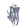


 PDBj
PDBj


 Assembly

 Spodoptera frugiperda (fall armyworm) / Strain (production host): Sf9 / References: UniProt: P0ABE7, UniProt: P47871
 Mass: 106.120 Da / Num. of mol.: 1 / Source method: obtained synthetically / Formula: C4H10O3
 Sample preparation
 / Beamline: 23-ID-D / Wavelength: 1.033 Å
 Processing