 Movie
Movie Controller

[English] 日本語
 Yorodumi
- PDB-4l41: Human Lactose synthase: A 2:1 complex between human alpha-lactalb... -
+ Open data

- Basic information
| Entry | Database: PDB / ID: 4l41 | ||||||
|---|---|---|---|---|---|---|---|
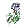
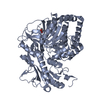
| Title | Human Lactose synthase: A 2:1 complex between human alpha-lactalbumin and human beta1,4-galactosyltransferase | ||||||
 Components Components |
| ||||||
 Keywords Keywords | Calcium Binding Protein/Transferase / GT-A fold / lactose synthase / Substrate binding / Golgi / Calcium Binding Protein-Transferase complex | ||||||
| Function / homology |  Function and homology information Function and homology informationDefective B4GALT1 causes CDG-2d / Interaction With Cumulus Cells And The Zona Pellucida / Defective B4GALT1 causes B4GALT1-CDG (CDG-2d) / Lactose synthesis / galactosyltransferase activity / Keratan sulfate biosynthesis / lactose synthase / neolactotriaosylceramide beta-1,4-galactosyltransferase / beta-N-acetylglucosaminylglycopeptide beta-1,4-galactosyltransferase / N-acetyllactosamine synthase ...Defective B4GALT1 causes CDG-2d / Interaction With Cumulus Cells And The Zona Pellucida / Defective B4GALT1 causes B4GALT1-CDG (CDG-2d) / Lactose synthesis / galactosyltransferase activity / Keratan sulfate biosynthesis / lactose synthase / neolactotriaosylceramide beta-1,4-galactosyltransferase / beta-N-acetylglucosaminylglycopeptide beta-1,4-galactosyltransferase / N-acetyllactosamine synthase / N-acetyllactosamine synthase activity / positive regulation of circulating fibrinogen levels / beta-N-acetylglucosaminylglycopeptide beta-1,4-galactosyltransferase activity / N-Glycan antennae elongation / penetration of zona pellucida / UDP-galactosyltransferase activity / regulation of acrosome reaction / Golgi trans cisterna / lactose synthase activity / lactose biosynthetic process / macrophage migration / oligosaccharide biosynthetic process / development of animal secondary sexual characteristics / desmosome / acute inflammatory response / galactose metabolic process / Pre-NOTCH Processing in Golgi / positive regulation of epithelial cell proliferation involved in wound healing / binding of sperm to zona pellucida / protein N-linked glycosylation / angiogenesis involved in wound healing / Transferases; Glycosyltransferases; Hexosyltransferases / Golgi cisterna membrane / azurophil granule membrane / epithelial cell development / enzyme-substrate adaptor activity / alpha-tubulin binding / beta-tubulin binding / extracellular matrix organization / epithelial cell proliferation / secretory granule membrane / filopodium / lipid metabolic process / brush border membrane / Golgi lumen / negative regulation of epithelial cell proliferation / lysozyme activity / manganese ion binding / cell-cell signaling / defense response to Gram-negative bacterium / basolateral plasma membrane / cell adhesion / defense response to bacterium / defense response to Gram-positive bacterium / positive regulation of apoptotic process / Golgi membrane / external side of plasma membrane / apoptotic process / calcium ion binding / Neutrophil degranulation / Golgi apparatus / signal transduction / protein-containing complex / : / extracellular exosome / membrane / identical protein binding / plasma membrane Similarity search - Function | ||||||
| Biological species |  Homo sapiens (human) Homo sapiens (human) | ||||||
| Method |  X-RAY DIFFRACTION / SYNCHROTRON / MOLECULAR REPLACEMENT / Resolution: 2.7 Å X-RAY DIFFRACTION / SYNCHROTRON / MOLECULAR REPLACEMENT / Resolution: 2.7 Å | ||||||
 Authors Authors | Ramakrishnan, B. / Qasba, P.K. | ||||||
 Citation Citation | Journal: To be Published Title: Crystal Structure of Human Lactose Synthase forms a Novel 1:2 Complex between beta1,4Galactosyltransferase 1 and alpha-Lactalbumin Proteins Authors: Ramakrishnan, B. / Qasba, P.K. | ||||||
| History |
|
- Structure visualization
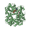
| Structure viewer | Molecule: MolmilJmol/JSmol |
|---|
- Downloads & links
-Download
| PDBx/mmCIF format | 4l41.cif.gz | 116.9 KB | Display | PDBx/mmCIF format |
|---|---|---|---|---|
| PDB format | pdb4l41.ent.gz | 90.2 KB | Display | PDB format |
| PDBx/mmJSON format | 4l41.json.gz | Tree view | PDBx/mmJSON format | |
| Others |  Other downloads Other downloads |
-Validation report
| Arichive directory | https://data.pdbj.org/pub/pdb/validation_reports/l4/4l41ftp://data.pdbj.org/pub/pdb/validation_reports/l4/4l41 | HTTPS FTP |
|---|
-Related structure data


-Links
 PDBj
PDBj


- Assembly
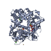
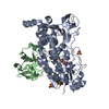
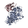
| Deposited unit | 
| ||||||||
|---|---|---|---|---|---|---|---|---|---|
| 1 |
| ||||||||
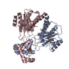
| Unit cell |
|
-Components
| #1: Protein | Mass: 14164.206 Da / Num. of mol.: 2 / Fragment: UNP residues 19-142 Source method: isolated from a genetically manipulated source Details: expressed with 11 amino acide N-terminal T7-tag / Source: (gene. exp.) Homo sapiens (human) / Gene: 4-galactosyltransferase, beta1, LALBA, LYZL7 / Plasmid: pET23a / Production host:  #2: Protein | | Mass: 32766.277 Da / Num. of mol.: 1 / Fragment: UNP residues 126-398 / Mutation: R337T, C338T Source method: isolated from a genetically manipulated source Details: contains an addition single amino acids Alanine at the N-terminal Source: (gene. exp.) Homo sapiens (human) / Gene: alpha-lactalbumin, B4GALT1, GGTB2 / Plasmid: pET17 / Production host: #3: Chemical |   Mass: 40.078 Da / Num. of mol.: 2 / Source method: obtained synthetically / Formula: Ca Mass: 40.078 Da / Num. of mol.: 2 / Source method: obtained synthetically / Formula: Ca#4: Chemical | ChemComp-SO4 / |   Mass: 96.063 Da / Num. of mol.: 1 / Source method: obtained synthetically / Formula: SO4 Mass: 96.063 Da / Num. of mol.: 1 / Source method: obtained synthetically / Formula: SO4#5: Water | ChemComp-HOH / |  Mass: 18.015 Da / Num. of mol.: 31 / Source method: isolated from a natural source / Formula: H2O Mass: 18.015 Da / Num. of mol.: 31 / Source method: isolated from a natural source / Formula: H2OHas protein modification | Y | |
|---|
-Experimental details
-Experiment
| Experiment | Method: X-RAY DIFFRACTION / Number of used crystals: 1 |
|---|
- Sample preparation
| Crystal | Density Matthews: 2.17 Å3/Da / Density % sol: 43.42 % |
|---|---|
| Crystal grow | Temperature: 298 K / Method: vapor diffusion, hanging drop / pH: 6.5 Details: 100 mM mes buffer pH 7.0, 12% PEG 4000, VAPOR DIFFUSION, HANGING DROP, temperature 298K |
-Data collection
| Diffraction | Mean temperature: 100 K |
|---|---|
| Diffraction source | Source: SYNCHROTRON / Site: NSLS  / Beamline: X9B / Wavelength: 1 Å / Beamline: X9B / Wavelength: 1 Å |
| Detector | Type: ADSC QUANTUM 4 / Detector: CCD / Date: Sep 2, 2000 / Details: mirrors |
| Radiation | Monochromator: graphite / Protocol: SINGLE WAVELENGTH / Monochromatic (M) / Laue (L): M / Scattering type: x-ray |
| Radiation wavelength | Wavelength: 1 Å / Relative weight: 1 |
| Reflection | Resolution: 2.7→50 Å / Num. all: 14802 / Num. obs: 14000 / % possible obs: 97.4 % / Observed criterion σ(I): 1 / Redundancy: 3 % / Rsym value: 0.11 / Net I/σ(I): 10.1 |
| Reflection shell | Resolution: 2.7→2.8 Å / Redundancy: 2.9 % / Mean I/σ(I) obs: 2.1 / Rsym value: 0.57 / % possible all: 98.4 |
- Processing
| Software |
| ||||||||||||||||||||||||||||||||||||||||||
|---|---|---|---|---|---|---|---|---|---|---|---|---|---|---|---|---|---|---|---|---|---|---|---|---|---|---|---|---|---|---|---|---|---|---|---|---|---|---|---|---|---|---|---|
| Refinement | Method to determine structure: MOLECULAR REPLACEMENT Starting model: PDB ENTRIES 1A4V AND 2AEC Resolution: 2.7→19.521 Å / SU ML: 0.24 / σ(F): 1.37 / Phase error: 23.49 / Stereochemistry target values: ML
| ||||||||||||||||||||||||||||||||||||||||||
| Solvent computation | Shrinkage radii: 0.9 Å / VDW probe radii: 1.11 Å / Solvent model: FLAT BULK SOLVENT MODEL | ||||||||||||||||||||||||||||||||||||||||||
| Refinement step | Cycle: LAST / Resolution: 2.7→19.521 Å
| ||||||||||||||||||||||||||||||||||||||||||
| Refine LS restraints |
| ||||||||||||||||||||||||||||||||||||||||||
| LS refinement shell |
|
