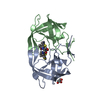
Mass: 10769.635 Da / Num. of mol.: 1 Source method: isolated from a genetically manipulated source Source: (gene. exp.) Human immunodeficiency virus 1 / Gene: pol / Production host: Escherichia coli (E. coli) / References: UniProt: Q000H7
#2: Protein
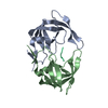
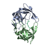
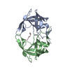
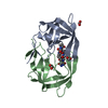
MDR769HIV-1protease
Mass: 10771.607 Da / Num. of mol.: 1 Source method: isolated from a genetically manipulated source Source: (gene. exp.) Human immunodeficiency virus 1 / Gene: pol / Production host: Escherichia coli (E. coli) / References: UniProt: Q000H7
Mass: 18.015 Da / Num. of mol.: 222 / Source method: isolated from a natural source / Formula: H2O
-
Experimental details
-
Experiment
Experiment
Method: X-RAY DIFFRACTION / Number of used crystals: 1
-
Sample preparation
Crystal
Density Matthews: 2.27 Å3/Da / Density % sol: 45.8 %
Crystal grow
Temperature: 298 K / Method: vapor diffusion, hanging drop / pH: 7.6 Details: The hanging drop vapor diffusion method was used to form the bipyramidal crystals of the MDR 769 protease. Using a grid screen consisting of sodium chloride (0.7 to 1.4 M) and MES-HEPES ...Details: The hanging drop vapor diffusion method was used to form the bipyramidal crystals of the MDR 769 protease. Using a grid screen consisting of sodium chloride (0.7 to 1.4 M) and MES-HEPES buffer (pH 5.5 to 8.1), the HIV-1 protease substrate complex crystals formed overnight at 22 C. Routinely, 0.2 mm crystals, in the longest dimension, were obtained after 14 days of incubation. In each well, there were two droplets, containing 1 lL of protease substrate mixture, 1 lL of reservoir solution and 2 lL of protease substrate mixture, 1 lL of reservoir solution, respectively, 07 mL of well solution., VAPOR DIFFUSION, HANGING DROP, temperature 298K
In the structure databanks used in Yorodumi, some data are registered as the other names, "COVID-19 virus" and "2019-nCoV". Here are the details of the virus and the list of structure data.
Jan 31, 2019. EMDB accession codes are about to change! (news from PDBe EMDB page)
EMDB accession codes are about to change! (news from PDBe EMDB page)
The allocation of 4 digits for EMDB accession codes will soon come to an end. Whilst these codes will remain in use, new EMDB accession codes will include an additional digit and will expand incrementally as the available range of codes is exhausted. The current 4-digit format prefixed with “EMD-” (i.e. EMD-XXXX) will advance to a 5-digit format (i.e. EMD-XXXXX), and so on. It is currently estimated that the 4-digit codes will be depleted around Spring 2019, at which point the 5-digit format will come into force.
The EM Navigator/Yorodumi systems omit the EMD- prefix.
Related info.:Q: What is EMD? / ID/Accession-code notation in Yorodumi/EM Navigator
Yorodumi is a browser for structure data from EMDB, PDB, SASBDB, etc.
This page is also the successor to EM Navigator detail page, and also detail information page/front-end page for Omokage search.
The word "yorodu" (or yorozu) is an old Japanese word meaning "ten thousand". "mi" (miru) is to see.
Related info.:EMDB / PDB / SASBDB / Comparison of 3 databanks / Yorodumi Search / Aug 31, 2016. New EM Navigator & Yorodumi / Yorodumi Papers / Jmol/JSmol / Function and homology information / Changes in new EM Navigator and Yorodumi
 Movie
Movie Controller

 Yorodumi
 Open data
 Basic information
 Components
 Keywords
 Function and homology information
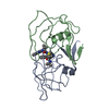
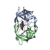
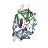
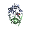
 Human immunodeficiency virus 1
 X-RAY DIFFRACTION /
  Authors
 Citation
 Structure visualization
 Downloads & links
 Other downloads
 PDBj
PDBj
 Assembly
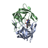

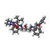
 Mass: 628.801 Da / Num. of mol.: 1 / Source method: obtained synthetically / Formula: C37H48N4O5
 Mass: 18.015 Da / Num. of mol.: 222 / Source method: isolated from a natural source / Formula: H2O
 Sample preparation
 / Beamline: 21-ID-D / Wavelength: 1 Å
 Processing