[English] 日本語
 Yorodumi
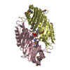
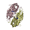
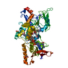
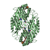
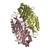
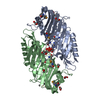
- PDB-4kso: Crystal Structure of Circadian clock protein KaiB from S.Elongatus -
+ Open data
- Basic information
| Entry | Database: PDB / ID: 4kso | ||||||
|---|---|---|---|---|---|---|---|
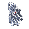
| Title | Crystal Structure of Circadian clock protein KaiB from S.Elongatus | ||||||
 Components Components | Circadian clock protein KaiB | ||||||
 Keywords Keywords | CIRCADIAN CLOCK PROTEIN / CYANOBACTERIAL CIRCADIAN CLOCK PROTEIN KaiB / CIRCADIAN CLOCK / KaiC Protein / Soluble | ||||||
| Function / homology |  Function and homology information Function and homology informationnegative regulation of phosphorylation / entrainment of circadian clock / circadian rhythm / regulation of circadian rhythm / identical protein binding / plasma membrane / cytoplasm Similarity search - Function | ||||||
| Biological species |  Synechococcus elongatus (bacteria) Synechococcus elongatus (bacteria) | ||||||
| Method |  X-RAY DIFFRACTION / X-RAY DIFFRACTION /  SYNCHROTRON / SYNCHROTRON /  MOLECULAR REPLACEMENT / Resolution: 2.622 Å MOLECULAR REPLACEMENT / Resolution: 2.622 Å | ||||||
 Authors Authors | Pattanayek, R. / Egli, M. | ||||||
 Citation Citation |  Journal: J Mol Biol / Year: 2013 Journal: J Mol Biol / Year: 2013Title: CryoEM and molecular dynamics of the circadian KaiB-KaiC complex indicates that KaiB monomers interact with KaiC and block ATP binding clefts. Authors: Seth A Villarreal / Rekha Pattanayek / Dewight R Williams / Tetsuya Mori / Ximing Qin / Carl H Johnson / Martin Egli / Phoebe L Stewart /  Abstract: The circadian control of cellular processes in cyanobacteria is regulated by a posttranslational oscillator formed by three Kai proteins. During the oscillator cycle, KaiA serves to promote ...The circadian control of cellular processes in cyanobacteria is regulated by a posttranslational oscillator formed by three Kai proteins. During the oscillator cycle, KaiA serves to promote autophosphorylation of KaiC while KaiB counteracts this effect. Here, we present a crystallographic structure of the wild-type Synechococcus elongatus KaiB and a cryo-electron microscopy (cryoEM) structure of a KaiBC complex. The crystal structure shows the expected dimer core structure and significant conformational variations of the KaiB C-terminal region, which is functionally important in maintaining rhythmicity. The KaiBC sample was formed with a C-terminally truncated form of KaiC, KaiC-Δ489, which is persistently phosphorylated. The KaiB-KaiC-Δ489 structure reveals that the KaiC hexamer can bind six monomers of KaiB, which form a continuous ring of density in the KaiBC complex. We performed cryoEM-guided molecular dynamics flexible fitting simulations with crystal structures of KaiB and KaiC to probe the KaiBC protein-protein interface. This analysis indicated a favorable binding mode for the KaiB monomer on the CII end of KaiC, involving two adjacent KaiC subunits and spanning an ATP binding cleft. A KaiC mutation, R468C, which has been shown to affect the affinity of KaiB for KaiC and lengthen the period in a bioluminescence rhythm assay, is found within the middle of the predicted KaiBC interface. The proposed KaiB binding mode blocks access to the ATP binding cleft in the CII ring of KaiC, which provides insight into how KaiB might influence the phosphorylation status of KaiC. | ||||||
| History |
|
- Structure visualization
| Structure viewer | Molecule:  Molmil Molmil Jmol/JSmol Jmol/JSmol |
|---|
- Downloads & links
- Download
| PDBx/mmCIF format |  4kso.cif.gz 4kso.cif.gz | 90.8 KB | Display |  PDBx/mmCIF format PDBx/mmCIF format |
|---|---|---|---|---|
| PDB format |  pdb4kso.ent.gz pdb4kso.ent.gz | 70.1 KB | Display |  PDB format PDB format |
| PDBx/mmJSON format |  4kso.json.gz 4kso.json.gz | Tree view |  PDBx/mmJSON format PDBx/mmJSON format | |
| Others |  Other downloads Other downloads |
-Validation report
| Arichive directory |  https://data.pdbj.org/pub/pdb/validation_reports/ks/4kso https://data.pdbj.org/pub/pdb/validation_reports/ks/4kso ftp://data.pdbj.org/pub/pdb/validation_reports/ks/4kso ftp://data.pdbj.org/pub/pdb/validation_reports/ks/4kso | HTTPS FTP |
|---|
-Related structure data
| Related structure data |  5672C  2qkeS S: Starting model for refinement C: citing same article ( |
|---|---|
| Similar structure data |
- Links
Links
- Assembly
| Deposited unit | 
| ||||||||
|---|---|---|---|---|---|---|---|---|---|
| 1 |
| ||||||||
| Unit cell |
|
- Components
| #1: Protein | Mass: 11450.387 Da / Num. of mol.: 4 Source method: isolated from a genetically manipulated source Source: (gene. exp.)  Synechococcus elongatus (bacteria) / Strain: PCC 7942 / Gene: kaiB / Plasmid: pGex vector / Production host: Synechococcus elongatus (bacteria) / Strain: PCC 7942 / Gene: kaiB / Plasmid: pGex vector / Production host:  #2: Water | ChemComp-HOH / | |
|---|
-Experimental details
-Experiment
| Experiment | Method:  X-RAY DIFFRACTION / Number of used crystals: 1 X-RAY DIFFRACTION / Number of used crystals: 1 |
|---|
- Sample preparation
| Crystal | Density Matthews: 2.33 Å3/Da / Density % sol: 47.23 % |
|---|---|
| Crystal grow | Temperature: 292 K / Method: vapor diffusion / pH: 5.7 Details: KaiB were grown from droplets containing 6.5 mg/mL KaiB-KaiC complex, 20 mM Tris (pH 7.8), 100 mM NaCl, 5 mM MgCl2,1 mM ATP and 2 mM BME. The reservoir solution was 100 mM sodium acetate, ...Details: KaiB were grown from droplets containing 6.5 mg/mL KaiB-KaiC complex, 20 mM Tris (pH 7.8), 100 mM NaCl, 5 mM MgCl2,1 mM ATP and 2 mM BME. The reservoir solution was 100 mM sodium acetate, 500 mM sodium formate and 5% glycerol (v/v), VAPOR DIFFUSION, temperature 292K |
-Data collection
| Diffraction | Mean temperature: 113 K |
|---|---|
| Diffraction source | Source:  SYNCHROTRON / Site: SYNCHROTRON / Site:  APS APS  / Beamline: 21-ID-G / Wavelength: 0.98 Å / Beamline: 21-ID-G / Wavelength: 0.98 Å |
| Detector | Type: MARMOSAIC 300 mm CCD / Detector: CCD / Date: Dec 14, 2012 / Details: Mirrors |
| Radiation | Monochromator: C(111) / Protocol: SINGLE WAVELENGTH / Monochromatic (M) / Laue (L): M / Scattering type: x-ray |
| Radiation wavelength | Wavelength: 0.98 Å / Relative weight: 1 |
| Reflection | Resolution: 2.6→30 Å / Num. obs: 12670 / % possible obs: 100 % / Observed criterion σ(F): 2 / Rmerge(I) obs: 0.06 / Net I/σ(I): 14.6 |
| Reflection shell | Resolution: 2.6→2.69 Å / Rmerge(I) obs: 0.267 / Mean I/σ(I) obs: 2.1 / % possible all: 99.9 |
- Processing
| Software |
| ||||||||||||||||||||||||||||
|---|---|---|---|---|---|---|---|---|---|---|---|---|---|---|---|---|---|---|---|---|---|---|---|---|---|---|---|---|---|
| Refinement | Method to determine structure:  MOLECULAR REPLACEMENT MOLECULAR REPLACEMENTStarting model: 2QKE Resolution: 2.622→29.689 Å / SU ML: 0.45 / σ(F): 1.39 / Phase error: 41.33 / Stereochemistry target values: ML
| ||||||||||||||||||||||||||||
| Solvent computation | Shrinkage radii: 0.9 Å / VDW probe radii: 1.11 Å / Solvent model: FLAT BULK SOLVENT MODEL | ||||||||||||||||||||||||||||
| Refinement step | Cycle: LAST / Resolution: 2.622→29.689 Å
| ||||||||||||||||||||||||||||
| Refine LS restraints |
| ||||||||||||||||||||||||||||
| LS refinement shell |
|
 Movie
Movie Controller



 PDBj
PDBj

