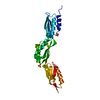
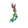
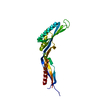
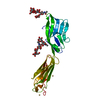
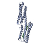
- PDB-4kqt: Crystal structure of a putative outer membrane chaperone (OmpH-li... -
+
Open data
ID or keywords:
Loading...
-
Basic information
Entry
Database: PDB / ID: 4kqt
Title
Crystal structure of a putative outer membrane chaperone (OmpH-like) (CC_1914) from Caulobacter crescentus CB15 at 2.83 A resolution (PSI Community Target, Shapiro)
Components
Putative outer membrane chaperone (OmpH-like)
Keywords
CHAPERONE / PF03938 family / Structural Genomics / Joint Center for Structural Genomics / JCSG / Protein Structure Initiative / PSI-BIOLOGY
Type: DECTRIS PILATUS 6M / Detector: PIXEL / Date: Jan 23, 2013 Details: Flat mirror (vertical focusing); single crystal Si(111) bent monochromator (horizontal focusing)
Radiation
Monochromator: single crystal Si(111) bent / Protocol: MAD / Monochromatic (M) / Laue (L): M / Scattering type: x-ray
Radiation wavelength
ID
Wavelength (Å)
Relative weight
1
0.97917
1
2
0.91837
1
3
0.97862
1
Reflection
Resolution: 2.83→45.677 Å / Num. obs: 9290 / % possible obs: 99.7 % / Observed criterion σ(I): -3 / Biso Wilson estimate: 90.539 Å2 / Rmerge(I) obs: 0.074 / Net I/σ(I): 16.65
Reflection shell
Resolution (Å)
Rmerge(I) obs
Mean I/σ(I) obs
Num. measured obs
Num. unique obs
Diffraction-ID
% possible all
2.83-2.93
0.946
1.9
5054
903
1
99.8
2.93-3.05
0.687
2.4
4935
934
1
99.6
3.05-3.19
0.395
4.2
5041
913
1
99.7
3.19-3.35
0.278
5.9
5119
886
1
99.9
3.35-3.56
0.159
10
5265
919
1
100
3.56-3.84
0.099
15.4
5264
946
1
99.9
3.84-4.22
0.067
21.1
4851
919
1
99.5
4.22-4.82
0.049
29
5271
913
1
100
4.82-6.05
0.049
27.4
4997
948
1
99.7
6.05-45.677
0.025
45.6
5266
1008
1
99.3
-
Phasing
Phasing
Method: MAD
-
Processing
Software
Name
Version
Classification
NB
MolProbity
3beta29
modelbuilding
PDB_EXTRACT
3.1
dataextraction
SHELX
phasing
SHARP
phasing
XSCALE
July4, 2012
datascaling
REFMAC
5.7.0032
refinement
XDS
datareduction
SHELXD
phasing
Refinement
Method to determine structure: MAD / Resolution: 2.83→45.677 Å / Cor.coef. Fo:Fc: 0.953 / Cor.coef. Fo:Fc free: 0.922 / Occupancy max: 1 / Occupancy min: 0.5 / SU B: 27.679 / SU ML: 0.235 / Cross valid method: THROUGHOUT / σ(F): 0 / ESU R: 0.371 / ESU R Free: 0.275 Stereochemistry target values: MAXIMUM LIKELIHOOD WITH PHASES Details: 1. A MET-INHIBITION PROTOCOL WAS USED FOR SELENOMETHIONINE INCORPORATION DURING PROTEIN EXPRESSION. THE OCCUPANCY OF THE SE ATOMS IN THE MSE RESIDUES WAS REDUCED TO 0.75 FOR THE REDUCED ...Details: 1. A MET-INHIBITION PROTOCOL WAS USED FOR SELENOMETHIONINE INCORPORATION DURING PROTEIN EXPRESSION. THE OCCUPANCY OF THE SE ATOMS IN THE MSE RESIDUES WAS REDUCED TO 0.75 FOR THE REDUCED SCATTERING POWER DUE TO PARTIAL S-MET INCORPORATION. 2. ATOM RECORD CONTAINS SUM OF TLS AND RESIDUAL B FACTORS. ANISOU RECORD CONTAINS SUM OF TLS AND RESIDUAL U FACTORS. 3. THE MAD PHASES WERE USED AS RESTRAINTS DURING REFINEMENT. 4. PEG MODELED IS PRESENT IN CRYO/CRYSTALLIZATION CONDITIONS. 5. HYDROGENS HAVE BEEN ADDED IN THE RIDING POSITIONS. 6. ZERO-OCCUPANCY ATOMS WERE PLACED NEAR THE THREE-FOLD AXIS TO EXCLUDE SOLVENT MASK NEAR THIS REGION. 7. WATERS WERE EXCLUDED FROM AUTOMATIC TLS ASSIGNMENT.
Rfactor
Num. reflection
% reflection
Selection details
Rfree
0.2452
441
4.7 %
RANDOM
Rwork
0.2084
-
-
-
obs
0.2101
8849
99.74 %
-
Solvent computation
Ion probe radii: 0.8 Å / Shrinkage radii: 0.8 Å / VDW probe radii: 1.2 Å / Solvent model: MASK
In the structure databanks used in Yorodumi, some data are registered as the other names, "COVID-19 virus" and "2019-nCoV". Here are the details of the virus and the list of structure data.
Jan 31, 2019. EMDB accession codes are about to change! (news from PDBe EMDB page)
EMDB accession codes are about to change! (news from PDBe EMDB page)
The allocation of 4 digits for EMDB accession codes will soon come to an end. Whilst these codes will remain in use, new EMDB accession codes will include an additional digit and will expand incrementally as the available range of codes is exhausted. The current 4-digit format prefixed with “EMD-” (i.e. EMD-XXXX) will advance to a 5-digit format (i.e. EMD-XXXXX), and so on. It is currently estimated that the 4-digit codes will be depleted around Spring 2019, at which point the 5-digit format will come into force.
The EM Navigator/Yorodumi systems omit the EMD- prefix.
Related info.:Q: What is EMD? / ID/Accession-code notation in Yorodumi/EM Navigator
Yorodumi is a browser for structure data from EMDB, PDB, SASBDB, etc.
This page is also the successor to EM Navigator detail page, and also detail information page/front-end page for Omokage search.
The word "yorodu" (or yorozu) is an old Japanese word meaning "ten thousand". "mi" (miru) is to see.
Related info.:EMDB / PDB / SASBDB / Comparison of 3 databanks / Yorodumi Search / Aug 31, 2016. New EM Navigator & Yorodumi / Yorodumi Papers / Jmol/JSmol / Function and homology information / Changes in new EM Navigator and Yorodumi
 Movie
Movie Controller

 Yorodumi
 Open data
 Basic information
 Components
 Keywords
 Function and homology information
 Caulobacter crescentus (bacteria)
 X-RAY DIFFRACTION /
  Authors
 Citation
 Structure visualization
 Downloads & links
 Other downloads


 PDBj
PDBj
 Assembly
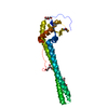

 Mass: 106.120 Da / Num. of mol.: 2 / Source method: obtained synthetically / Formula: C4H10O3
 Mass: 150.173 Da / Num. of mol.: 2 / Source method: obtained synthetically / Formula: C6H14O4
 Mass: 18.015 Da / Num. of mol.: 4 / Source method: isolated from a natural source / Formula: H2O
 Sample preparation
 / Beamline: BL11-1 / Wavelength: 0.97917,0.91837,0.97862
 Processing