 Movie
Movie Controller

[English] 日本語
 Yorodumi
- PDB-4kic: Crystal structure of methyltransferase from Streptomyces hygrosco... -
+ Open data

- Basic information
| Entry | Database: PDB / ID: 4kic | |||||||||
|---|---|---|---|---|---|---|---|---|---|---|
| Title | Crystal structure of methyltransferase from Streptomyces hygroscopicus complexed with S-adenosyl-L-methionine and phenylpyruvic acid | |||||||||
 Components Components | Methyltransferase MppJ | |||||||||
 Keywords Keywords | TRANSFERASE / Rossmann Fold / Methyltransferase / SAM Binding PPY Binding | |||||||||
| Function / homology |  Function and homology information Function and homology informationphenylpyruvate C3-methyltransferase / antibiotic biosynthetic process / methyltransferase activity / methylation Similarity search - Function | |||||||||
| Biological species |  Streptomyces hygroscopicus (bacteria) Streptomyces hygroscopicus (bacteria) | |||||||||
| Method |  X-RAY DIFFRACTION / MOLECULAR REPLACEMENT / Resolution: 2.43 Å X-RAY DIFFRACTION / MOLECULAR REPLACEMENT / Resolution: 2.43 Å | |||||||||
 Authors Authors | Liu, Y.C. / Zou, X.W. / Chan, H.C. / Huang, C.J. / Li, T.L. | |||||||||
 Citation Citation | Journal: Acta Crystallogr.,Sect.D / Year: 2014 Title: Structure and mechanism of a nonhaem-iron SAM-dependent C-methyltransferase and its engineering to a hydratase and an O-methyltransferase Authors: Zou, X.W. / Liu, Y.C. / Hsu, N.S. / Huang, C.J. / Lyu, S.Y. / Chan, H.C. / Chang, C.Y. / Yeh, H.W. / Lin, K.H. / Wu, C.J. / Tsai, M.D. / Li, T.L. | |||||||||
| History |
|
- Structure visualization
| Structure viewer | Molecule: MolmilJmol/JSmol |
|---|
- Downloads & links
-Download
| PDBx/mmCIF format | 4kic.cif.gz | 155.2 KB | Display | PDBx/mmCIF format |
|---|---|---|---|---|
| PDB format | pdb4kic.ent.gz | 119.2 KB | Display | PDB format |
| PDBx/mmJSON format | 4kic.json.gz | Tree view | PDBx/mmJSON format | |
| Others |  Other downloads Other downloads |
-Validation report
| Arichive directory | https://data.pdbj.org/pub/pdb/validation_reports/ki/4kicftp://data.pdbj.org/pub/pdb/validation_reports/ki/4kic | HTTPS FTP |
|---|
-Related structure data
| Related structure data |  4kibSC  4kifC  4kigC  4m6xC  4m6yC  4m71C  4m72C  4m73C  4m74C S: Starting model for refinement C: citing same article ( |
|---|---|
| Similar structure data |


-Links
 PDBj
PDBj
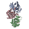
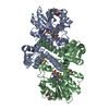
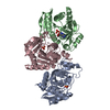
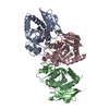
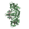
- Assembly
| Deposited unit | 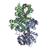
| ||||||||
|---|---|---|---|---|---|---|---|---|---|
| 1 |
| ||||||||
| Unit cell |
|
-Components
-Protein , 1 types, 2 molecules AB
| #1: Protein | Mass: 39400.289 Da / Num. of mol.: 2 Source method: isolated from a genetically manipulated source Source: (gene. exp.) Streptomyces hygroscopicus (bacteria) / Gene: mppJ / Production host: |
|---|
-Non-polymers , 7 types, 321 molecules 


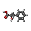


| #2: Chemical |  Mass: 398.437 Da / Num. of mol.: 2 / Source method: obtained synthetically / Formula: C15H22N6O5S Mass: 398.437 Da / Num. of mol.: 2 / Source method: obtained synthetically / Formula: C15H22N6O5S#3: Chemical |  Mass: 164.158 Da / Num. of mol.: 2 / Source method: obtained synthetically / Formula: C9H8O3 Mass: 164.158 Da / Num. of mol.: 2 / Source method: obtained synthetically / Formula: C9H8O3#4: Chemical |  Mass: 55.845 Da / Num. of mol.: 2 / Source method: obtained synthetically / Formula: Fe Mass: 55.845 Da / Num. of mol.: 2 / Source method: obtained synthetically / Formula: Fe#5: Chemical |  Type: peptide-like / Mass: 166.174 Da / Num. of mol.: 2 / Source method: obtained synthetically / Formula: C9H10O3 Type: peptide-like / Mass: 166.174 Da / Num. of mol.: 2 / Source method: obtained synthetically / Formula: C9H10O3#6: Chemical | ChemComp-CA /  Mass: 40.078 Da / Num. of mol.: 13 / Source method: obtained synthetically / Formula: Ca Mass: 40.078 Da / Num. of mol.: 13 / Source method: obtained synthetically / Formula: Ca#7: Chemical | ChemComp-IOD / |  Mass: 126.904 Da / Num. of mol.: 1 / Source method: obtained synthetically / Formula: I Mass: 126.904 Da / Num. of mol.: 1 / Source method: obtained synthetically / Formula: I#8: Water | ChemComp-HOH / | Mass: 18.015 Da / Num. of mol.: 299 / Source method: isolated from a natural source / Formula: H2O |
|---|
-Experimental details
-Experiment
| Experiment | Method: X-RAY DIFFRACTION / Number of used crystals: 1 |
|---|
- Sample preparation
| Crystal | Density Matthews: 2.43 Å3/Da / Density % sol: 49.34 % |
|---|---|
| Crystal grow | Temperature: 293 K / Method: vapor diffusion, hanging drop / pH: 7.5 Details: 16% PEG3350, 0.2M sodium iodide, pH 7.5, VAPOR DIFFUSION, HANGING DROP, temperature 293K |
-Data collection
| Diffraction | Mean temperature: 100 K |
|---|---|
| Diffraction source | Source: ROTATING ANODE / Type: RIGAKU / Wavelength: 1.54 Å |
| Detector | Type: RIGAKU RAXIS IV / Detector: IMAGE PLATE / Date: Sep 24, 2010 |
| Radiation | Monochromator: Diffracted Beam Monochromator / Protocol: SINGLE WAVELENGTH / Monochromatic (M) / Laue (L): M / Scattering type: x-ray |
| Radiation wavelength | Wavelength: 1.54 Å / Relative weight: 1 |
| Reflection | Resolution: 2.43→19 Å / Num. all: 27511 / Num. obs: 27511 / % possible obs: 97.1 % / Observed criterion σ(F): 2 / Observed criterion σ(I): 2 |
| Reflection shell | Resolution: 2.43→2.54 Å / % possible all: 90.4 |
- Processing
| Software |
| ||||||||||||||||||||||||||||||||||||||||||||||||||||||||||||
|---|---|---|---|---|---|---|---|---|---|---|---|---|---|---|---|---|---|---|---|---|---|---|---|---|---|---|---|---|---|---|---|---|---|---|---|---|---|---|---|---|---|---|---|---|---|---|---|---|---|---|---|---|---|---|---|---|---|---|---|---|---|
| Refinement | Method to determine structure: MOLECULAR REPLACEMENT Starting model: PDB ENTRY 4KIB Resolution: 2.43→19 Å / Cor.coef. Fo:Fc: 0.963 / Cor.coef. Fo:Fc free: 0.923 / SU B: 8.33 / SU ML: 0.19 / Cross valid method: THROUGHOUT / σ(F): 2 / ESU R: 0.437 / ESU R Free: 0.267 / Stereochemistry target values: MAXIMUM LIKELIHOOD / Details: HYDROGENS HAVE BEEN ADDED IN THE RIDING POSITIONS
| ||||||||||||||||||||||||||||||||||||||||||||||||||||||||||||
| Solvent computation | Ion probe radii: 0.8 Å / Shrinkage radii: 0.8 Å / VDW probe radii: 1.2 Å / Solvent model: MASK | ||||||||||||||||||||||||||||||||||||||||||||||||||||||||||||
| Displacement parameters | Biso mean: 42.589 Å2
| ||||||||||||||||||||||||||||||||||||||||||||||||||||||||||||
| Refinement step | Cycle: LAST / Resolution: 2.43→19 Å
| ||||||||||||||||||||||||||||||||||||||||||||||||||||||||||||
| Refine LS restraints |
| ||||||||||||||||||||||||||||||||||||||||||||||||||||||||||||
| LS refinement shell | Resolution: 2.43→2.488 Å / Total num. of bins used: 20
|
