 Movie
Movie Controller

[English] 日本語
 Yorodumi
- PDB-5lc1: L-threonine dehydrogenase from Trypanosoma brucei with NAD and th... -
+ Open data

- Basic information
| Entry | Database: PDB / ID: 5lc1 | |||||||||
|---|---|---|---|---|---|---|---|---|---|---|
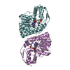
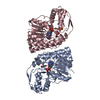
| Title | L-threonine dehydrogenase from Trypanosoma brucei with NAD and the inhibitor pyruvate bound. | |||||||||
 Components Components | L-threonine 3-dehydrogenase | |||||||||
 Keywords Keywords | OXIDOREDUCTASE / Dehydrogenase / holo-enzyme / Rossman fold | |||||||||
| Function / homology |  Function and homology information Function and homology informationL-threonine 3-dehydrogenase / L-threonine 3-dehydrogenase activity / L-threonine catabolic process / nucleotide binding Similarity search - Function | |||||||||
| Biological species |  | |||||||||
| Method |  X-RAY DIFFRACTION / SYNCHROTRON / MOLECULAR REPLACEMENT / Resolution: 2.1 Å X-RAY DIFFRACTION / SYNCHROTRON / MOLECULAR REPLACEMENT / Resolution: 2.1 Å | |||||||||
 Authors Authors | Erskine, P.T. / Adjogatse, E. / Wood, S.P. / Cooper, J.B. | |||||||||
 Citation Citation | Journal: Acta Crystallogr D Struct Biol / Year: 2018 Title: Structure and function of L-threonine-3-dehydrogenase from the parasitic protozoan Trypanosoma brucei revealed by X-ray crystallography and geometric simulations. Authors: Adjogatse, E. / Erskine, P. / Wells, S.A. / Kelly, J.M. / Wilden, J.D. / Chan, A.W.E. / Selwood, D. / Coker, A. / Wood, S. / Cooper, J.B. | |||||||||
| History |
|
- Structure visualization
| Structure viewer | Molecule: MolmilJmol/JSmol |
|---|
- Downloads & links
-Download
| PDBx/mmCIF format | 5lc1.cif.gz | 431.6 KB | Display | PDBx/mmCIF format |
|---|---|---|---|---|
| PDB format | pdb5lc1.ent.gz | 351.7 KB | Display | PDB format |
| PDBx/mmJSON format | 5lc1.json.gz | Tree view | PDBx/mmJSON format | |
| Others |  Other downloads Other downloads |
-Validation report
| Arichive directory | https://data.pdbj.org/pub/pdb/validation_reports/lc/5lc1ftp://data.pdbj.org/pub/pdb/validation_reports/lc/5lc1 | HTTPS FTP |
|---|
-Related structure data
| Related structure data |  5k4qC 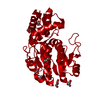 5k4tC  5k4uC  5k4vC  5k4wC  5k4yC  5k50C  5l9aSC S: Starting model for refinement C: citing same article ( |
|---|---|
| Similar structure data |


-Links
 PDBj
PDBj


- Assembly
| Deposited unit | 
| ||||||||
|---|---|---|---|---|---|---|---|---|---|
| 1 | 
| ||||||||
| 2 | 
| ||||||||
| 3 | 
| ||||||||
| 4 | 
| ||||||||
| 5 | 
| ||||||||
| 6 | 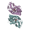
| ||||||||
| Unit cell |
|
-Components
-Protein , 1 types, 6 molecules ABCDEF
| #1: Protein | Mass: 35847.305 Da / Num. of mol.: 6 Source method: isolated from a genetically manipulated source Source: (gene. exp.)  |
|---|
-Non-polymers , 6 types, 1985 molecules 


| #2: Chemical | ChemComp-NAD /  Mass: 663.425 Da / Num. of mol.: 6 / Source method: obtained synthetically / Formula: C21H27N7O14P2 / Comment: NAD*YM Mass: 663.425 Da / Num. of mol.: 6 / Source method: obtained synthetically / Formula: C21H27N7O14P2 / Comment: NAD*YM#3: Chemical | ChemComp-PYR /  Mass: 88.062 Da / Num. of mol.: 6 / Source method: obtained synthetically / Formula: C3H4O3 Mass: 88.062 Da / Num. of mol.: 6 / Source method: obtained synthetically / Formula: C3H4O3#4: Chemical |  Mass: 78.133 Da / Num. of mol.: 2 / Source method: obtained synthetically / Formula: C2H6OS Mass: 78.133 Da / Num. of mol.: 2 / Source method: obtained synthetically / Formula: C2H6OS#5: Chemical |  Mass: 59.044 Da / Num. of mol.: 3 / Source method: obtained synthetically / Formula: C2H3O2 Mass: 59.044 Da / Num. of mol.: 3 / Source method: obtained synthetically / Formula: C2H3O2#6: Chemical | ChemComp-NA /  Mass: 22.990 Da / Num. of mol.: 6 / Source method: obtained synthetically / Formula: Na Mass: 22.990 Da / Num. of mol.: 6 / Source method: obtained synthetically / Formula: Na#7: Water | ChemComp-HOH / | Mass: 18.015 Da / Num. of mol.: 1962 / Source method: isolated from a natural source / Formula: H2O |
|---|
-Experimental details
-Experiment
| Experiment | Method: X-RAY DIFFRACTION / Number of used crystals: 1 |
|---|
- Sample preparation
| Crystal | Density Matthews: 2.2 Å3/Da / Density % sol: 45.1 % |
|---|---|
| Crystal grow | Temperature: 293 K / Method: vapor diffusion, hanging drop / pH: 7.5 Details: 0.1 M HEPES pH 7.5, 20 % w/v PEG 10K; TDH 2.0 mg/ml, 1 mM NAD(+), 30 mM pyruvate. |
-Data collection
| Diffraction | Mean temperature: 100 K |
|---|---|
| Diffraction source | Source: SYNCHROTRON / Site: ESRF  / Beamline: ID29 / Wavelength: 1.074 Å / Beamline: ID29 / Wavelength: 1.074 Å |
| Detector | Type: ADSC QUANTUM 315r / Detector: CCD / Date: Nov 11, 2009 |
| Radiation | Protocol: SINGLE WAVELENGTH / Monochromatic (M) / Laue (L): M / Scattering type: x-ray |
| Radiation wavelength | Wavelength: 1.074 Å / Relative weight: 1 |
| Reflection | Resolution: 2.1→95.5 Å / Num. obs: 117204 / % possible obs: 97.3 % / Observed criterion σ(F): 0 / Observed criterion σ(I): 0 / Redundancy: 7.5 % / Biso Wilson estimate: 21.2 Å2 / Rmerge(I) obs: 0.145 / Net I/σ(I): 11.4 |
| Reflection shell | Resolution: 2.1→2.2 Å / Redundancy: 3.3 % / Rmerge(I) obs: 0.525 / Mean I/σ(I) obs: 3.2 / % possible all: 81.9 |
- Processing
| Software |
| ||||||||||||||||||||||||||||||||||||||||||||||||||||||||||||||||||||||||||||||||||||||||||||||||||||||||||||||||||||||||||||||||||||||||||||||||||||||||||||||||||||||||||||||||||||||
|---|---|---|---|---|---|---|---|---|---|---|---|---|---|---|---|---|---|---|---|---|---|---|---|---|---|---|---|---|---|---|---|---|---|---|---|---|---|---|---|---|---|---|---|---|---|---|---|---|---|---|---|---|---|---|---|---|---|---|---|---|---|---|---|---|---|---|---|---|---|---|---|---|---|---|---|---|---|---|---|---|---|---|---|---|---|---|---|---|---|---|---|---|---|---|---|---|---|---|---|---|---|---|---|---|---|---|---|---|---|---|---|---|---|---|---|---|---|---|---|---|---|---|---|---|---|---|---|---|---|---|---|---|---|---|---|---|---|---|---|---|---|---|---|---|---|---|---|---|---|---|---|---|---|---|---|---|---|---|---|---|---|---|---|---|---|---|---|---|---|---|---|---|---|---|---|---|---|---|---|---|---|---|---|
| Refinement | Method to determine structure: MOLECULAR REPLACEMENT Starting model: 5L9A Resolution: 2.1→95.5 Å / Cor.coef. Fo:Fc: 0.968 / Cor.coef. Fo:Fc free: 0.935 / SU B: 4.668 / SU ML: 0.122 / Cross valid method: THROUGHOUT / ESU R: 0.202 / ESU R Free: 0.176 Details: HYDROGENS HAVE BEEN ADDED IN THE RIDING POSITIONS. Note that the R-free set was chosen in thin resolution shells and hence the outer shell (Shell 1, below) did not contain any free-set ...Details: HYDROGENS HAVE BEEN ADDED IN THE RIDING POSITIONS. Note that the R-free set was chosen in thin resolution shells and hence the outer shell (Shell 1, below) did not contain any free-set reflections. The R-free and N(R-free) given below relate the highest resolution shell which contained free-set reflections.
| ||||||||||||||||||||||||||||||||||||||||||||||||||||||||||||||||||||||||||||||||||||||||||||||||||||||||||||||||||||||||||||||||||||||||||||||||||||||||||||||||||||||||||||||||||||||
| Solvent computation | Ion probe radii: 0.8 Å / Shrinkage radii: 0.8 Å / VDW probe radii: 1.2 Å | ||||||||||||||||||||||||||||||||||||||||||||||||||||||||||||||||||||||||||||||||||||||||||||||||||||||||||||||||||||||||||||||||||||||||||||||||||||||||||||||||||||||||||||||||||||||
| Displacement parameters | Biso mean: 22.767 Å2
| ||||||||||||||||||||||||||||||||||||||||||||||||||||||||||||||||||||||||||||||||||||||||||||||||||||||||||||||||||||||||||||||||||||||||||||||||||||||||||||||||||||||||||||||||||||||
| Refinement step | Cycle: 1 / Resolution: 2.1→95.5 Å
| ||||||||||||||||||||||||||||||||||||||||||||||||||||||||||||||||||||||||||||||||||||||||||||||||||||||||||||||||||||||||||||||||||||||||||||||||||||||||||||||||||||||||||||||||||||||
| Refine LS restraints |
|