 Movie
Movie Controller

[English] 日本語
 Yorodumi
- PDB-5k4u: Three-dimensional structure of L-threonine 3-dehydrogenase from T... -
+ Open data

- Basic information
| Entry | Database: PDB / ID: 5k4u | ||||||
|---|---|---|---|---|---|---|---|
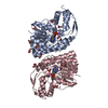
| Title | Three-dimensional structure of L-threonine 3-dehydrogenase from Trypanosoma brucei showing different active site loop conformations between dimer subunits, refined to 1.9 angstroms | ||||||
 Components Components | L-threonine 3-dehydrogenase | ||||||
 Keywords Keywords | OXIDOREDUCTASE / short-chain dehydrogenase / ligand-bound / holo | ||||||
| Function / homology |  Function and homology information Function and homology informationL-threonine 3-dehydrogenase / L-threonine 3-dehydrogenase activity / L-threonine catabolic process / nucleotide binding Similarity search - Function | ||||||
| Biological species |  | ||||||
| Method |  X-RAY DIFFRACTION / SYNCHROTRON / MOLECULAR REPLACEMENT / Resolution: 1.9 Å X-RAY DIFFRACTION / SYNCHROTRON / MOLECULAR REPLACEMENT / Resolution: 1.9 Å | ||||||
 Authors Authors | Adjogatse, E.K. / Cooper, J.B. / Erskine, P.T. | ||||||
 Citation Citation | Journal: Acta Crystallogr D Struct Biol / Year: 2018 Title: Structure and function of L-threonine-3-dehydrogenase from the parasitic protozoan Trypanosoma brucei revealed by X-ray crystallography and geometric simulations. Authors: Adjogatse, E. / Erskine, P. / Wells, S.A. / Kelly, J.M. / Wilden, J.D. / Chan, A.W.E. / Selwood, D. / Coker, A. / Wood, S. / Cooper, J.B. | ||||||
| History |
|
- Structure visualization
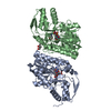
| Structure viewer | Molecule: MolmilJmol/JSmol |
|---|
- Downloads & links
-Download
| PDBx/mmCIF format | 5k4u.cif.gz | 144.3 KB | Display | PDBx/mmCIF format |
|---|---|---|---|---|
| PDB format | pdb5k4u.ent.gz | 112.1 KB | Display | PDB format |
| PDBx/mmJSON format | 5k4u.json.gz | Tree view | PDBx/mmJSON format | |
| Others |  Other downloads Other downloads |
-Validation report
| Arichive directory | https://data.pdbj.org/pub/pdb/validation_reports/k4/5k4uftp://data.pdbj.org/pub/pdb/validation_reports/k4/5k4u | HTTPS FTP |
|---|
-Related structure data
| Related structure data |  5k4qC  5k4tC  5k4vC  5k4wC  5k4yC  5k50C  5l9aC  5lc1C C: citing same article ( |
|---|---|
| Similar structure data |


-Links
 PDBj
PDBj- Assembly
| Deposited unit | 
| ||||||||
|---|---|---|---|---|---|---|---|---|---|
| 1 |
| ||||||||
| Unit cell |
|
-Components
| #1: Protein | Mass: 35709.160 Da / Num. of mol.: 2 Source method: isolated from a genetically manipulated source Source: (gene. exp.)  #2: Chemical |   Mass: 663.425 Da / Num. of mol.: 2 / Source method: obtained synthetically / Formula: C21H27N7O14P2 / Comment: NAD*YM Mass: 663.425 Da / Num. of mol.: 2 / Source method: obtained synthetically / Formula: C21H27N7O14P2 / Comment: NAD*YM#3: Chemical | ChemComp-GOL /   Mass: 92.094 Da / Num. of mol.: 5 / Source method: obtained synthetically / Formula: C3H8O3 Mass: 92.094 Da / Num. of mol.: 5 / Source method: obtained synthetically / Formula: C3H8O3#4: Chemical | ChemComp-ACT /   Mass: 59.044 Da / Num. of mol.: 5 / Source method: obtained synthetically / Formula: C2H3O2 Mass: 59.044 Da / Num. of mol.: 5 / Source method: obtained synthetically / Formula: C2H3O2#5: Water | ChemComp-HOH / |  Mass: 18.015 Da / Num. of mol.: 228 / Source method: isolated from a natural source / Formula: H2O Mass: 18.015 Da / Num. of mol.: 228 / Source method: isolated from a natural source / Formula: H2O |
|---|
-Experimental details
-Experiment
| Experiment | Method: X-RAY DIFFRACTION / Number of used crystals: 1 |
|---|
- Sample preparation
| Crystal | Density Matthews: 2.19 Å3/Da / Density % sol: 43.94 % |
|---|---|
| Crystal grow | Temperature: 298 K / Method: vapor diffusion, hanging drop / pH: 5.6 Details: 0.1M tri-sodium citrate, 30% w/v PEG 4K, 0.2M sodium acetate, NAD+ (8.5mM) |
-Data collection
| Diffraction | Mean temperature: 100 K | |||||||||||||||
|---|---|---|---|---|---|---|---|---|---|---|---|---|---|---|---|---|
| Diffraction source | Source: SYNCHROTRON / Site: Diamond  / Beamline: I04-1 / Wavelength: 0.9173 Å / Beamline: I04-1 / Wavelength: 0.9173 Å | |||||||||||||||
| Detector | Type: DECTRIS PILATUS 2M / Detector: PIXEL / Date: Feb 19, 2012 | |||||||||||||||
| Radiation | Protocol: SINGLE WAVELENGTH / Monochromatic (M) / Laue (L): M / Scattering type: x-ray | |||||||||||||||
| Radiation wavelength | Wavelength: 0.9173 Å / Relative weight: 1 | |||||||||||||||
| Reflection | Resolution: 1.79→29.29 Å / Num. obs: 61881 / % possible obs: 98 % / Redundancy: 3.3 % / Rmerge(I) obs: 0.074 / Net I/σ(I): 12.7 | |||||||||||||||
| Reflection shell |
|
- Processing
| Software |
| |||||||||||||||||||||||||||||||||||||||||||||||||||||||||||||||||||||||||||
|---|---|---|---|---|---|---|---|---|---|---|---|---|---|---|---|---|---|---|---|---|---|---|---|---|---|---|---|---|---|---|---|---|---|---|---|---|---|---|---|---|---|---|---|---|---|---|---|---|---|---|---|---|---|---|---|---|---|---|---|---|---|---|---|---|---|---|---|---|---|---|---|---|---|---|---|---|
| Refinement | Method to determine structure: MOLECULAR REPLACEMENT / Resolution: 1.9→28.55 Å / Cor.coef. Fo:Fc: 0.95 / Cor.coef. Fo:Fc free: 0.923 / SU B: 3.027 / SU ML: 0.092 / Cross valid method: THROUGHOUT / σ(F): 0 / ESU R: 0.146 / ESU R Free: 0.143
| |||||||||||||||||||||||||||||||||||||||||||||||||||||||||||||||||||||||||||
| Solvent computation | Ion probe radii: 0.8 Å / Shrinkage radii: 0.8 Å / VDW probe radii: 1.2 Å | |||||||||||||||||||||||||||||||||||||||||||||||||||||||||||||||||||||||||||
| Displacement parameters | Biso max: 78.08 Å2 / Biso mean: 23.973 Å2 / Biso min: 9.25 Å2
| |||||||||||||||||||||||||||||||||||||||||||||||||||||||||||||||||||||||||||
| Refinement step | Cycle: final / Resolution: 1.9→28.55 Å
| |||||||||||||||||||||||||||||||||||||||||||||||||||||||||||||||||||||||||||
| Refine LS restraints |
| |||||||||||||||||||||||||||||||||||||||||||||||||||||||||||||||||||||||||||
| LS refinement shell | Resolution: 1.9→1.949 Å / Total num. of bins used: 20
|
