Type: MAR scanner 345 mm plate / Detector: IMAGE PLATE
Radiation
Monochromator: Ni FILTER / Protocol: SINGLE WAVELENGTH / Monochromatic (M) / Laue (L): M / Scattering type: x-ray
Radiation wavelength
Wavelength: 0.9793 Å / Relative weight: 1
Reflection
Resolution: 2.003→38.03 Å
-
Processing
Software
Name
Version
Classification
PHENIX
1.8.2_1309
refinement
MOLREP
phasing
REFMAC
5.6.0117
refinement
HKL-2000
datareduction
SCALA
datascaling
HKL-2000
datacollection
Refinement
Method to determine structure: MOLECULAR REPLACEMENT / Resolution: 2.003→38.03 Å / Cor.coef. Fo:Fc: 0.928 / Cor.coef. Fo:Fc free: 0.904 / SU ML: 0.2 / σ(F): 1.34 / Phase error: 22.84 / Stereochemistry target values: ML / Details: HYDROGENS HAVE BEEN USED IF PRESENT IN THE INPUT
Rfactor
Num. reflection
% reflection
Rfree
0.233
752
5.01 %
Rwork
0.1892
-
-
obs
0.1913
15024
98.82 %
Solvent computation
Shrinkage radii: 0.9 Å / VDW probe radii: 1.11 Å / Solvent model: FLAT BULK SOLVENT MODEL
Displacement parameters
Biso mean: 21.321 Å2
Baniso -1
Baniso -2
Baniso -3
1-
1.56 Å2
0 Å2
0.56 Å2
2-
-
-0.67 Å2
0 Å2
3-
-
-
-0.8 Å2
Refinement step
Cycle: LAST / Resolution: 2.003→38.03 Å
Protein
Nucleic acid
Ligand
Solvent
Total
Num. atoms
1559
0
26
72
1657
Refine LS restraints
Refine-ID
Type
Dev ideal
Number
X-RAY DIFFRACTION
f_bond_d
0.007
1626
X-RAY DIFFRACTION
f_angle_d
1.096
2213
X-RAY DIFFRACTION
f_dihedral_angle_d
13.677
574
X-RAY DIFFRACTION
f_chiral_restr
0.074
248
X-RAY DIFFRACTION
f_plane_restr
0.006
287
LS refinement shell
Resolution (Å)
Rfactor Rfree
Num. reflection Rfree
Rfactor Rwork
Num. reflection Rwork
Refine-ID
% reflection obs (%)
2.003-2.1576
0.2497
148
0.1841
2829
X-RAY DIFFRACTION
99
2.1576-2.3747
0.2497
155
0.177
2837
X-RAY DIFFRACTION
99
2.3747-2.7182
0.2403
145
0.1948
2860
X-RAY DIFFRACTION
99
2.7182-3.4243
0.2376
151
0.1932
2850
X-RAY DIFFRACTION
99
3.4243-38.0369
0.2175
153
0.1902
2896
X-RAY DIFFRACTION
98
+
About Yorodumi
-
News
-
Feb 9, 2022. New format data for meta-information of EMDB entries
New format data for meta-information of EMDB entries
Version 3 of the EMDB header file is now the official format.
The previous official version 1.9 will be removed from the archive.
In the structure databanks used in Yorodumi, some data are registered as the other names, "COVID-19 virus" and "2019-nCoV". Here are the details of the virus and the list of structure data.
Jan 31, 2019. EMDB accession codes are about to change! (news from PDBe EMDB page)
EMDB accession codes are about to change! (news from PDBe EMDB page)
The allocation of 4 digits for EMDB accession codes will soon come to an end. Whilst these codes will remain in use, new EMDB accession codes will include an additional digit and will expand incrementally as the available range of codes is exhausted. The current 4-digit format prefixed with “EMD-” (i.e. EMD-XXXX) will advance to a 5-digit format (i.e. EMD-XXXXX), and so on. It is currently estimated that the 4-digit codes will be depleted around Spring 2019, at which point the 5-digit format will come into force.
The EM Navigator/Yorodumi systems omit the EMD- prefix.
Related info.:Q: What is EMD? / ID/Accession-code notation in Yorodumi/EM Navigator
Yorodumi is a browser for structure data from EMDB, PDB, SASBDB, etc.
This page is also the successor to EM Navigator detail page, and also detail information page/front-end page for Omokage search.
The word "yorodu" (or yorozu) is an old Japanese word meaning "ten thousand". "mi" (miru) is to see.
Related info.:EMDB / PDB / SASBDB / Comparison of 3 databanks / Yorodumi Search / Aug 31, 2016. New EM Navigator & Yorodumi / Yorodumi Papers / Jmol/JSmol / Function and homology information / Changes in new EM Navigator and Yorodumi
 Movie
Movie Controller


 Open data
 Basic information
 Components
 Keywords
 Function and homology information
 X-RAY DIFFRACTION /
  Authors
 Citation
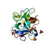
 Structure visualization
 Downloads & links
 Other downloads
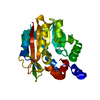

 PDBj
PDBj Assembly
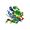
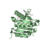
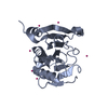
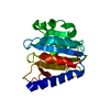
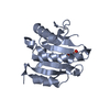
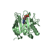
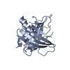
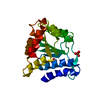
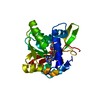

 Type: L-peptide linking / Mass: 384.411 Da / Num. of mol.: 1 / Source method: obtained synthetically / Formula: C14H20N6O5S
 Mass: 18.015 Da / Num. of mol.: 72 / Source method: isolated from a natural source / Formula: H2O
 Sample preparation
 Processing