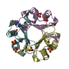
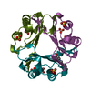
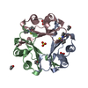
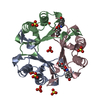
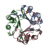
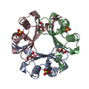
- PDB-4k9g: 1.55 A Crystal Structure of Macrophage Migration Inhibitory Facto... -
+
Open data
ID or keywords:
Loading...
-
Basic information
Entry
Database: PDB / ID: 4k9g
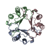
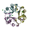
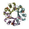
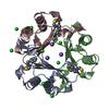
Title
1.55 A Crystal Structure of Macrophage Migration Inhibitory Factor bound to ISO-66 and a related compound
Components
Macrophage migration inhibitory factor
Keywords
ISOMERASE / cytokine / secreted/endocytosed
Function / homology
Function and homology information
: / positive regulation of myeloid leukocyte cytokine production involved in immune response / phenylpyruvate tautomerase / L-dopachrome isomerase / regulation of macrophage activation / dopachrome isomerase activity / phenylpyruvate tautomerase activity / cytokine receptor binding / negative regulation of myeloid cell apoptotic process / negative regulation of mature B cell apoptotic process ...: / positive regulation of myeloid leukocyte cytokine production involved in immune response / phenylpyruvate tautomerase / L-dopachrome isomerase / regulation of macrophage activation / dopachrome isomerase activity / phenylpyruvate tautomerase activity / cytokine receptor binding / negative regulation of myeloid cell apoptotic process / negative regulation of mature B cell apoptotic process / negative regulation of macrophage chemotaxis / carboxylic acid metabolic process / positive regulation of arachidonate secretion / positive regulation of lipopolysaccharide-mediated signaling pathway / prostaglandin biosynthetic process / positive regulation of chemokine (C-X-C motif) ligand 2 production / negative regulation of protein metabolic process / negative regulation of intrinsic apoptotic signaling pathway in response to DNA damage by p53 class mediator / protein homotrimerization / chemoattractant activity / negative regulation of DNA damage response, signal transduction by p53 class mediator / negative regulation of cellular senescence / positive regulation of cAMP/PKA signal transduction / positive regulation of phosphorylation / positive regulation of B cell proliferation / Gene and protein expression by JAK-STAT signaling after Interleukin-12 stimulation / negative regulation of cell migration / cytokine activity / positive regulation of cytokine production / Cell surface interactions at the vascular wall / DNA damage response, signal transduction by p53 class mediator / positive regulation of fibroblast proliferation / cellular senescence / positive regulation of tumor necrosis factor production / protease binding / secretory granule lumen / vesicle / ficolin-1-rich granule lumen / positive regulation of ERK1 and ERK2 cascade / cell surface receptor signaling pathway / inflammatory response / negative regulation of gene expression / innate immune response / positive regulation of cell population proliferation / Neutrophil degranulation / negative regulation of apoptotic process / cell surface / : / extracellular exosome / extracellular region / nucleoplasm / identical protein binding / plasma membrane / cytosol / cytoplasm Similarity search - Function
Mass: 18.015 Da / Num. of mol.: 281 / Source method: isolated from a natural source / Formula: H2O
-
Experimental details
-
Experiment
Experiment
Method: X-RAY DIFFRACTION / Number of used crystals: 1
-
Sample preparation
Crystal
Density Matthews: 3.71 Å3/Da / Density % sol: 66.85 %
Crystal grow
Temperature: 293 K / pH: 7.5 Details: 1.1 mM (14 mg/ml) MIF , 10 mM inhibitor in 10% DMSO, 18 mM NaCl, 18 mM tris(hydroxymethyl)aminomethane mixed 1:1 with reservoir containing 2 M ammonium sulfate, 0.1 M tris(hydroxymethyl) ...Details: 1.1 mM (14 mg/ml) MIF , 10 mM inhibitor in 10% DMSO, 18 mM NaCl, 18 mM tris(hydroxymethyl)aminomethane mixed 1:1 with reservoir containing 2 M ammonium sulfate, 0.1 M tris(hydroxymethyl)aminomethane, 3% isopropanol, pH 7.5, VAPOR DIFFUSION, HANGING DROP, temperature 293K
Resolution: 1.55→82.76 Å / Isotropic thermal model: isotropic / Cross valid method: THROUGHOUT / σ(F): 0 / Stereochemistry target values: ENGH & HUBER Details: REFINEMENT WAS BEGUN WITH CNS. REFMAC WAS USED IN LATER STAGES. IN ACTIVE SITE A, TWO FORMS OF THE INHIBITOR (RING-OPENED AND RING-CLOSED) WERE OBSERVED. THE LOWER OCCUPANCY FORM (RING- ...Details: REFINEMENT WAS BEGUN WITH CNS. REFMAC WAS USED IN LATER STAGES. IN ACTIVE SITE A, TWO FORMS OF THE INHIBITOR (RING-OPENED AND RING-CLOSED) WERE OBSERVED. THE LOWER OCCUPANCY FORM (RING-CLOSED) WAS OBSERVED IN MAPS OMITTING THIS INHIBITOR FORM WHEN CNS WAS USED FOR MAP CALCULATION, AND MISSING REFLECTIONS WERE NOT REPLACED BY FC. MAPS CALCULATED USING REFMAC, AS WELL AS CNS MAPS IN WHICH MISSING REFLECTIONS WERE REPLACED BY FC, DID NOT REVEAL THE LOWER OCCUPANCY INHIBITOR. TLS WAS USED IN LATER REFINEMENT STEPS. THE FINAL R-FACTORS LISTED TAKE THE TLS MODEL INTO ACCOUNT. WITHOUT TLS, R(OBS)=0.22629, R(WORK)=0.22590, R(FREE)=0.24575.
Rfactor
Num. reflection
% reflection
Selection details
Rfree
0.22
1597
-
RANDOM
Rwork
0.199
-
-
-
obs
0.199
78325
99.6 %
-
Solvent computation
Ion probe radii: 0.8 Å / Shrinkage radii: 0.8 Å / VDW probe radii: 1.2 Å / Solvent model: MASK
In the structure databanks used in Yorodumi, some data are registered as the other names, "COVID-19 virus" and "2019-nCoV". Here are the details of the virus and the list of structure data.
Jan 31, 2019. EMDB accession codes are about to change! (news from PDBe EMDB page)
EMDB accession codes are about to change! (news from PDBe EMDB page)
The allocation of 4 digits for EMDB accession codes will soon come to an end. Whilst these codes will remain in use, new EMDB accession codes will include an additional digit and will expand incrementally as the available range of codes is exhausted. The current 4-digit format prefixed with “EMD-” (i.e. EMD-XXXX) will advance to a 5-digit format (i.e. EMD-XXXXX), and so on. It is currently estimated that the 4-digit codes will be depleted around Spring 2019, at which point the 5-digit format will come into force.
The EM Navigator/Yorodumi systems omit the EMD- prefix.
Related info.:Q: What is EMD? / ID/Accession-code notation in Yorodumi/EM Navigator
Yorodumi is a browser for structure data from EMDB, PDB, SASBDB, etc.
This page is also the successor to EM Navigator detail page, and also detail information page/front-end page for Omokage search.
The word "yorodu" (or yorozu) is an old Japanese word meaning "ten thousand". "mi" (miru) is to see.
Related info.:EMDB / PDB / SASBDB / Comparison of 3 databanks / Yorodumi Search / Aug 31, 2016. New EM Navigator & Yorodumi / Yorodumi Papers / Jmol/JSmol / Function and homology information / Changes in new EM Navigator and Yorodumi
 Movie
Movie Controller

 Yorodumi
 Open data
 Basic information
 Components
 Keywords
 Function and homology information
 Homo sapiens (human)
 X-RAY DIFFRACTION /
  Authors
 Citation
 Structure visualization
 Downloads & links
 Other downloads


 PDBj
PDBj


 Assembly
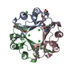

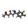
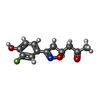


 Mass: 239.243 Da / Num. of mol.: 3 / Source method: obtained synthetically / Formula: C12H14FNO3
 Mass: 237.227 Da / Num. of mol.: 1 / Source method: obtained synthetically / Formula: C12H12FNO3
 Mass: 35.453 Da / Num. of mol.: 6 / Source method: obtained synthetically / Formula: Cl
 Mass: 96.063 Da / Num. of mol.: 1 / Source method: obtained synthetically / Formula: SO4
 Sample preparation
 / Beamline: X29A / Wavelength: 1.0809
  Processing