 Movie
Movie Controller


+ Open data

- Basic information
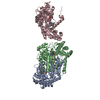
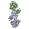
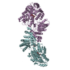
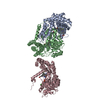
| Entry | Database: PDB / ID: 4k24 | |||||||||
|---|---|---|---|---|---|---|---|---|---|---|
| Title | Structure of anti-uPAR Fab ATN-658 in complex with uPAR | |||||||||
 Components Components |
| |||||||||
 Keywords Keywords | IMMUNE SYSTEM | |||||||||
| Function / homology |  Function and homology information Function and homology informationurokinase plasminogen activator receptor activity / Attachment of GPI anchor to uPAR / symbiont-containing vacuole / smooth muscle cell-matrix adhesion / peptidase inhibitor complex / rough endoplasmic reticulum lumen / positive regulation of integrin-mediated signaling pathway / u-plasminogen activator / regulation of smooth muscle cell-matrix adhesion / urokinase plasminogen activator signaling pathway ...urokinase plasminogen activator receptor activity / Attachment of GPI anchor to uPAR / symbiont-containing vacuole / smooth muscle cell-matrix adhesion / peptidase inhibitor complex / rough endoplasmic reticulum lumen / positive regulation of integrin-mediated signaling pathway / u-plasminogen activator / regulation of smooth muscle cell-matrix adhesion / urokinase plasminogen activator signaling pathway / regulation of plasminogen activation / positive regulation of homotypic cell-cell adhesion / regulation of integrin-mediated signaling pathway / alphav-beta3 integrin-vitronectin complex / protein complex involved in cell-matrix adhesion / regulation of fibrinolysis / regulation of wound healing / negative regulation of plasminogen activation / positive regulation of cell-substrate adhesion / positive regulation of vascular endothelial growth factor signaling pathway / serine-type endopeptidase complex / regulation of smooth muscle cell migration / Dissolution of Fibrin Clot / regulation of cell adhesion mediated by integrin / extracellular matrix binding / positive regulation of vascular endothelial growth factor receptor signaling pathway / protein localization to cell surface / scavenger receptor activity / extrinsic component of membrane / cell adhesion mediated by integrin / positive regulation of DNA binding / smooth muscle cell migration / Molecules associated with elastic fibres / plasminogen activation / positive regulation of smooth muscle cell migration / extracellular matrix structural constituent / Syndecan interactions / endodermal cell differentiation / positive regulation of wound healing / negative regulation of intrinsic apoptotic signaling pathway / oligodendrocyte differentiation / positive regulation of release of cytochrome c from mitochondria / polysaccharide binding / tertiary granule membrane / basement membrane / regulation of proteolysis / negative regulation of blood coagulation / negative regulation of fibrinolysis / protein polymerization / ECM proteoglycans / Integrin cell surface interactions / regulation of cell adhesion / serine protease inhibitor complex / specific granule membrane / fibrinolysis / positive regulation of epidermal growth factor receptor signaling pathway / collagen binding / negative regulation of proteolysis / cell projection / extracellular matrix organization / liver regeneration / Regulation of Complement cascade / integrin-mediated signaling pathway / cell-matrix adhesion / Developmental Lineage of Pancreatic Ductal Cells / positive regulation of protein phosphorylation / positive regulation of receptor-mediated endocytosis / Golgi lumen / integrin binding / chemotaxis / blood coagulation / regulation of cell population proliferation / cell migration / heparin binding / extracellular matrix / signaling receptor activity / blood microparticle / response to hypoxia / cell adhesion / immune response / positive regulation of cell migration / endoplasmic reticulum lumen / receptor ligand activity / serine-type endopeptidase activity / external side of plasma membrane / protein domain specific binding / signaling receptor binding / focal adhesion / Neutrophil degranulation / negative regulation of apoptotic process / endoplasmic reticulum membrane / enzyme binding / cell surface / signal transduction / endoplasmic reticulum / proteolysis / : / extracellular exosome / extracellular region / membrane Similarity search - Function | |||||||||
| Biological species |  Homo sapiens (human) Homo sapiens (human) | |||||||||
| Method |  X-RAY DIFFRACTION / SYNCHROTRON / MOLECULAR REPLACEMENT / Resolution: 4.5 Å X-RAY DIFFRACTION / SYNCHROTRON / MOLECULAR REPLACEMENT / Resolution: 4.5 Å | |||||||||
 Authors Authors | Huang, M.D. / Xu, X. / Yuan, C. | |||||||||
 Citation Citation | Journal: Plos One / Year: 2014 Title: Identification of a New Epitope in uPAR as a Target for the Cancer Therapeutic Monoclonal Antibody ATN-658, a Structural Homolog of the uPAR Binding Integrin CD11b ( alpha M) Authors: Xu, X. / Cai, Y. / Wei, Y. / Donate, F. / Juarez, J. / Parry, G. / Chen, L. / Meehan, E.J. / Ahn, R.W. / Ugolkov, A. / Dubrovskyi, O. / O'halloran, T.V. / Huang, M. / Mazar, A.P. | |||||||||
| History |
|
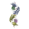
- Structure visualization
| Structure viewer | Molecule: MolmilJmol/JSmol |
|---|
- Downloads & links
-Download
| PDBx/mmCIF format | 4k24.cif.gz | 373.7 KB | Display | PDBx/mmCIF format |
|---|---|---|---|---|
| PDB format | pdb4k24.ent.gz | 306.9 KB | Display | PDB format |
| PDBx/mmJSON format | 4k24.json.gz | Tree view | PDBx/mmJSON format | |
| Others |  Other downloads Other downloads |
-Validation report
| Arichive directory | https://data.pdbj.org/pub/pdb/validation_reports/k2/4k24ftp://data.pdbj.org/pub/pdb/validation_reports/k2/4k24 | HTTPS FTP |
|---|
-Related structure data
| Related structure data |  4k23SC  3bt2S S: Starting model for refinement C: citing same article ( |
|---|---|
| Similar structure data |


-Links
 PDBj
PDBj


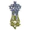
- Assembly
| Deposited unit | 
| ||||||||
|---|---|---|---|---|---|---|---|---|---|
| 1 | 
| ||||||||
| Unit cell |
|
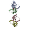
-Components
-Protein , 2 types, 2 molecules AU
| #1: Protein | Mass: 15359.318 Da / Num. of mol.: 1 / Fragment: UNP residues 21-153 Source method: isolated from a genetically manipulated source Source: (gene. exp.) Homo sapiens (human) / Gene: PLAU / Plasmid: pMT/Bip / Production host:  |
|---|---|
| #5: Protein | Mass: 31601.479 Da / Num. of mol.: 1 / Fragment: UNP residues 23-303 Source method: isolated from a genetically manipulated source Source: (gene. exp.) Homo sapiens (human) / Gene: MO3, PLAUR, UPAR / Plasmid: pMT/Bip / Production host: |
-Protein/peptide , 1 types, 1 molecules B
| #2: Protein/peptide | Mass: 4573.103 Da / Num. of mol.: 1 / Fragment: UNP residues 21-60 Source method: isolated from a genetically manipulated source Source: (gene. exp.) Homo sapiens (human) / Gene: VTN / Production host:  |
|---|
-Antibody , 2 types, 2 molecules HL
| #3: Antibody | Mass: 24482.459 Da / Num. of mol.: 1 / Source method: isolated from a natural source / Source: (natural) |
|---|---|
| #4: Antibody | Mass: 24156.930 Da / Num. of mol.: 1 / Source method: isolated from a natural source / Source: (natural) |
-Sugars , 3 types, 4 molecules 

| #6: Polysaccharide | Source method: isolated from a genetically manipulated source #7: Sugar | ChemComp-MAN / |  Type: D-saccharide, alpha linking / Mass: 180.156 Da / Num. of mol.: 1 Type: D-saccharide, alpha linking / Mass: 180.156 Da / Num. of mol.: 1Source method: isolated from a genetically manipulated source Formula: C6H12O6 #8: Sugar | ChemComp-NAG / |  Type: D-saccharide, beta linking / Mass: 221.208 Da / Num. of mol.: 1 Type: D-saccharide, beta linking / Mass: 221.208 Da / Num. of mol.: 1Source method: isolated from a genetically manipulated source Formula: C8H15NO6 |
|---|
-Details
| Has protein modification | Y |
|---|---|
| Sequence details | A SEQUENCE DATABASE REFERENCE FOR ENTITY 3 AND 4 DOES NOT CURRENTLY EXIST. |
-Experimental details
-Experiment
| Experiment | Method: X-RAY DIFFRACTION / Number of used crystals: 1 |
|---|
- Sample preparation
| Crystal | Density Matthews: 5.1 Å3/Da / Density % sol: 75.88 % |
|---|---|
| Crystal grow | Temperature: 295 K / Method: vapor diffusion, sitting drop / pH: 7.5 Details: 0.1M HEPES pH 7.5, 55%(v/v) Tacsimate, 2%(v/v) 2-methyl-1,3-propanediol, vapor diffusion, sitting drop, temperature 295K |
-Data collection
| Diffraction | Mean temperature: 100 K | |||||||||||||||||||||||||||||||||||||||||||||||||||||||
|---|---|---|---|---|---|---|---|---|---|---|---|---|---|---|---|---|---|---|---|---|---|---|---|---|---|---|---|---|---|---|---|---|---|---|---|---|---|---|---|---|---|---|---|---|---|---|---|---|---|---|---|---|---|---|---|---|
| Diffraction source | Source: SYNCHROTRON / Site: NSLS  / Beamline: X29A / Wavelength: 1.04 Å / Beamline: X29A / Wavelength: 1.04 Å | |||||||||||||||||||||||||||||||||||||||||||||||||||||||
| Detector | Type: MARMOSAIC 300 mm CCD / Detector: CCD / Date: Jan 3, 2007 | |||||||||||||||||||||||||||||||||||||||||||||||||||||||
| Radiation | Protocol: SINGLE WAVELENGTH / Monochromatic (M) / Laue (L): M / Scattering type: x-ray | |||||||||||||||||||||||||||||||||||||||||||||||||||||||
| Radiation wavelength | Wavelength: 1.04 Å / Relative weight: 1 | |||||||||||||||||||||||||||||||||||||||||||||||||||||||
| Reflection | Resolution: 4.5→27 Å / Num. obs: 12451 / % possible obs: 99.7 % / Redundancy: 4.4 % / Rmerge(I) obs: 0.139 / Net I/σ(I): 7.9 | |||||||||||||||||||||||||||||||||||||||||||||||||||||||
| Reflection shell |
|
- Processing
| Software |
| ||||||||||||||||||||||||||||||||||||||||||||||||||||||||||||||||||||||||||||||||||||||||||||||||||||||||||||||||||||||||||||||||||||||||||||||||||||||
|---|---|---|---|---|---|---|---|---|---|---|---|---|---|---|---|---|---|---|---|---|---|---|---|---|---|---|---|---|---|---|---|---|---|---|---|---|---|---|---|---|---|---|---|---|---|---|---|---|---|---|---|---|---|---|---|---|---|---|---|---|---|---|---|---|---|---|---|---|---|---|---|---|---|---|---|---|---|---|---|---|---|---|---|---|---|---|---|---|---|---|---|---|---|---|---|---|---|---|---|---|---|---|---|---|---|---|---|---|---|---|---|---|---|---|---|---|---|---|---|---|---|---|---|---|---|---|---|---|---|---|---|---|---|---|---|---|---|---|---|---|---|---|---|---|---|---|---|---|---|---|---|
| Refinement | Method to determine structure: MOLECULAR REPLACEMENT Starting model: 3BT2(chain A, B, U), 4K23(chain H, L) Resolution: 4.5→26.902 Å / Occupancy max: 1 / Occupancy min: 1 / SU ML: 0.51 / σ(F): 1.34 / Phase error: 30.96 / Stereochemistry target values: ML
| ||||||||||||||||||||||||||||||||||||||||||||||||||||||||||||||||||||||||||||||||||||||||||||||||||||||||||||||||||||||||||||||||||||||||||||||||||||||
| Solvent computation | Shrinkage radii: 0.9 Å / VDW probe radii: 1.11 Å / Solvent model: FLAT BULK SOLVENT MODEL | ||||||||||||||||||||||||||||||||||||||||||||||||||||||||||||||||||||||||||||||||||||||||||||||||||||||||||||||||||||||||||||||||||||||||||||||||||||||
| Displacement parameters | Biso mean: 284.9089 Å2
| ||||||||||||||||||||||||||||||||||||||||||||||||||||||||||||||||||||||||||||||||||||||||||||||||||||||||||||||||||||||||||||||||||||||||||||||||||||||
| Refinement step | Cycle: LAST / Resolution: 4.5→26.902 Å
| ||||||||||||||||||||||||||||||||||||||||||||||||||||||||||||||||||||||||||||||||||||||||||||||||||||||||||||||||||||||||||||||||||||||||||||||||||||||
| Refine LS restraints |
| ||||||||||||||||||||||||||||||||||||||||||||||||||||||||||||||||||||||||||||||||||||||||||||||||||||||||||||||||||||||||||||||||||||||||||||||||||||||
| LS refinement shell |
| ||||||||||||||||||||||||||||||||||||||||||||||||||||||||||||||||||||||||||||||||||||||||||||||||||||||||||||||||||||||||||||||||||||||||||||||||||||||
| Refinement TLS params. | Method: refined / Refine-ID: X-RAY DIFFRACTION
| ||||||||||||||||||||||||||||||||||||||||||||||||||||||||||||||||||||||||||||||||||||||||||||||||||||||||||||||||||||||||||||||||||||||||||||||||||||||
| Refinement TLS group |
|
