 Movie
Movie Controller

[English] 日本語
 Yorodumi
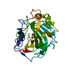
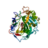
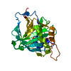
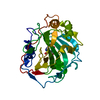
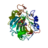
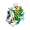
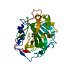
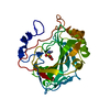
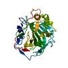
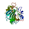
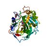
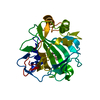
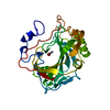
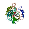
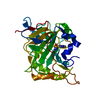
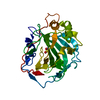
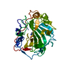
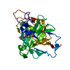
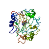
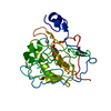
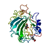
- PDB-4k13: Structure of HCAIX mimic (HCAII with 5 mutations in active site) ... -
+ Open data

- Basic information
| Entry | Database: PDB / ID: 4k13 | ||||||
|---|---|---|---|---|---|---|---|
| Title | Structure of HCAIX mimic (HCAII with 5 mutations in active site) in complex with dorzolamide | ||||||
 Components Components | Carbonic anhydrase 2 | ||||||
 Keywords Keywords | LYASE / alpha beta fold | ||||||
| Function / homology |  Function and homology information Function and homology information: / positive regulation of dipeptide transmembrane transport / secretion / regulation of monoatomic anion transport / cyanamide hydratase / cyanamide hydratase activity / arylesterase activity / regulation of chloride transport / Reversible hydration of carbon dioxide / positive regulation of synaptic transmission, GABAergic ...: / positive regulation of dipeptide transmembrane transport / secretion / regulation of monoatomic anion transport / cyanamide hydratase / cyanamide hydratase activity / arylesterase activity / regulation of chloride transport / Reversible hydration of carbon dioxide / positive regulation of synaptic transmission, GABAergic / morphogenesis of an epithelium / angiotensin-activated signaling pathway / Developmental Lineage of Pancreatic Ductal Cells / carbonic anhydrase / regulation of intracellular pH / carbonate dehydratase activity / neuron cellular homeostasis / carbon dioxide transport / Erythrocytes take up oxygen and release carbon dioxide / Erythrocytes take up carbon dioxide and release oxygen / apical part of cell / myelin sheath / extracellular exosome / zinc ion binding / plasma membrane / cytosol / cytoplasm Similarity search - Function | ||||||
| Biological species |  Homo sapiens (human) Homo sapiens (human) | ||||||
| Method |  X-RAY DIFFRACTION / MOLECULAR REPLACEMENT / Resolution: 1.6 Å X-RAY DIFFRACTION / MOLECULAR REPLACEMENT / Resolution: 1.6 Å | ||||||
 Authors Authors | Biswas, S. / McKenna, R. | ||||||
 Citation Citation | Journal: To be Published Title: Developing isoform specific inhibitors for hCAIX using CAIX mimic Authors: Biswas, S. / West, D. / Pinard, M. / McKenna, R. | ||||||
| History |
|
- Structure visualization
| Structure viewer | Molecule: MolmilJmol/JSmol |
|---|
- Downloads & links
-Download
| PDBx/mmCIF format | 4k13.cif.gz | 75.4 KB | Display | PDBx/mmCIF format |
|---|---|---|---|---|
| PDB format | pdb4k13.ent.gz | 53.9 KB | Display | PDB format |
| PDBx/mmJSON format | 4k13.json.gz | Tree view | PDBx/mmJSON format | |
| Others |  Other downloads Other downloads |
-Validation report
| Arichive directory | https://data.pdbj.org/pub/pdb/validation_reports/k1/4k13ftp://data.pdbj.org/pub/pdb/validation_reports/k1/4k13 | HTTPS FTP |
|---|
-Related structure data
| Related structure data | 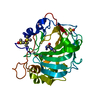 4k0zC 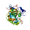 4k1qC 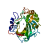 2iliS C: citing same article ( S: Starting model for refinement |
|---|---|
| Similar structure data |

-Links
 PDBj
PDBj

- Assembly
| Deposited unit | 
| ||||||||
|---|---|---|---|---|---|---|---|---|---|
| 1 |
| ||||||||
| Unit cell |
|
-Components
| #1: Protein | Mass: 28900.574 Da / Num. of mol.: 1 / Mutation: A65S, N67Q, E69T, F131L, K170E Source method: isolated from a genetically manipulated source Source: (gene. exp.) Homo sapiens (human) / Gene: CA2 / Production host:  | ||||
|---|---|---|---|---|---|
| #2: Chemical | ChemComp-ZN /   Mass: 65.409 Da / Num. of mol.: 1 / Source method: obtained synthetically / Formula: Zn Mass: 65.409 Da / Num. of mol.: 1 / Source method: obtained synthetically / Formula: Zn | ||||
| #3: Chemical |   Mass: 78.133 Da / Num. of mol.: 2 / Source method: obtained synthetically / Formula: C2H6OS / Comment: DMSO, precipitant*YM Mass: 78.133 Da / Num. of mol.: 2 / Source method: obtained synthetically / Formula: C2H6OS / Comment: DMSO, precipitant*YM#4: Chemical | ChemComp-ETS / ( | 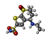  Mass: 324.440 Da / Num. of mol.: 1 / Source method: obtained synthetically / Formula: C10H16N2O4S3 / Comment: medication*YM Mass: 324.440 Da / Num. of mol.: 1 / Source method: obtained synthetically / Formula: C10H16N2O4S3 / Comment: medication*YM#5: Water | ChemComp-HOH / |  Mass: 18.015 Da / Num. of mol.: 311 / Source method: isolated from a natural source / Formula: H2O Mass: 18.015 Da / Num. of mol.: 311 / Source method: isolated from a natural source / Formula: H2O |
-Experimental details
-Experiment
| Experiment | Method: X-RAY DIFFRACTION / Number of used crystals: 1 |
|---|
- Sample preparation
| Crystal | Density Matthews: 2.11 Å3/Da / Density % sol: 41.73 % |
|---|---|
| Crystal grow | Temperature: 298 K / Method: vapor diffusion, hanging drop / pH: 8 Details: 1.6 M sodium citrate, 50 mM Tris, pH 8.0, VAPOR DIFFUSION, HANGING DROP, temperature 298K |
-Data collection
| Diffraction | Mean temperature: 100 K |
|---|---|
| Diffraction source | Source: ROTATING ANODE / Type: RIGAKU RUH3R / Wavelength: 1.54 Å |
| Detector | Type: RIGAKU RAXIS IV++ / Detector: IMAGE PLATE / Date: Feb 22, 2012 |
| Radiation | Monochromator: varimax optics / Protocol: SINGLE WAVELENGTH / Monochromatic (M) / Laue (L): M / Scattering type: x-ray |
| Radiation wavelength | Wavelength: 1.54 Å / Relative weight: 1 |
| Reflection | Resolution: 1.6→50 Å / Num. all: 24187 / Num. obs: 24187 / % possible obs: 75.4 % / Observed criterion σ(F): 0 / Observed criterion σ(I): -3 / Redundancy: 1.8 % / Rmerge(I) obs: 0.048 / Rsym value: 0.048 / Net I/σ(I): 13.1 |
| Reflection shell | Resolution: 1.6→1.66 Å / Redundancy: 1.8 % / Rmerge(I) obs: 0.289 / Mean I/σ(I) obs: 3.6 / Rsym value: 0.289 / % possible all: 72.1 |
- Processing
| Software |
| ||||||||||||||||||||||||||||||||
|---|---|---|---|---|---|---|---|---|---|---|---|---|---|---|---|---|---|---|---|---|---|---|---|---|---|---|---|---|---|---|---|---|---|
| Refinement | Method to determine structure: MOLECULAR REPLACEMENT Starting model: PDB ENTRY 2ILI Resolution: 1.6→30.473 Å / Occupancy max: 1 / Occupancy min: 0 / SU ML: 0.15 / σ(F): 1.37 / Phase error: 19.83 / Stereochemistry target values: Engh & Huber
| ||||||||||||||||||||||||||||||||
| Solvent computation | Shrinkage radii: 0.9 Å / VDW probe radii: 1.11 Å / Solvent model: FLAT BULK SOLVENT MODEL | ||||||||||||||||||||||||||||||||
| Displacement parameters | Biso max: 64.82 Å2 / Biso mean: 19.2151 Å2 / Biso min: 4.98 Å2 | ||||||||||||||||||||||||||||||||
| Refinement step | Cycle: LAST / Resolution: 1.6→30.473 Å
| ||||||||||||||||||||||||||||||||
| Refine LS restraints |
|
