[English] 日本語
Yorodumi
- PDB-6ymb: MicroED structure of human carbonic anhydrase II -

+
Open data


ID or keywords:

Loading...

-
Basic information

Entry
Database: PDB / ID: 6ymb
TitleMicroED structure of human carbonic anhydrase II
Componentscarbonic anhydrase 2
KeywordsLYASE / carbonic anhydrase / MicroED
Function / homology
Function and homology information


positive regulation of cellular pH reduction / positive regulation of dipeptide transmembrane transport / regulation of monoatomic anion transport / secretion / cyanamide hydratase / cyanamide hydratase activity / arylesterase activity / regulation of chloride transport / Reversible hydration of carbon dioxide / angiotensin-activated signaling pathway ...positive regulation of cellular pH reduction / positive regulation of dipeptide transmembrane transport / regulation of monoatomic anion transport / secretion / cyanamide hydratase / cyanamide hydratase activity / arylesterase activity / regulation of chloride transport / Reversible hydration of carbon dioxide / angiotensin-activated signaling pathway / positive regulation of synaptic transmission, GABAergic / morphogenesis of an epithelium / regulation of intracellular pH / carbonic anhydrase / carbonate dehydratase activity / carbon dioxide transport / Erythrocytes take up oxygen and release carbon dioxide / Erythrocytes take up carbon dioxide and release oxygen / neuron cellular homeostasis / one-carbon metabolic process / apical part of cell / myelin sheath / zinc ion binding / extracellular exosome / plasma membrane / cytoplasm / cytosol
Similarity search - Function
Carbonic anhydrase, alpha-class, conserved site / Alpha-carbonic anhydrases signature. / Carbonic anhydrase, alpha-class / Alpha carbonic anhydrase domain / Alpha carbonic anhydrase domain superfamily / Eukaryotic-type carbonic anhydrase / Alpha-carbonic anhydrases profile. / Eukaryotic-type carbonic anhydrase
Similarity search - Domain/homology
Carbonic anhydrase 2
Similarity search - Component
Biological speciesHomo sapiens (human)
MethodELECTRON CRYSTALLOGRAPHY / electron crystallography / MOLECULAR REPLACEMENT / cryo EM / Resolution: 2.5 Å
AuthorsClabbers, M.T.B. / Fisher, S.Z. / Coincon, M. / Zou, X. / Xu, H.
Funding support Sweden, 3items
OrganizationGrant numberCountry
Swedish Research Council2017-05333 Sweden
Swedish Research Council2019-00815 Sweden
Knut and Alice Wallenberg Foundation2018.0237 Sweden
Citation
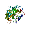
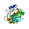
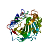
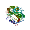
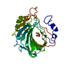
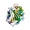
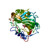
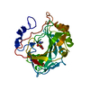
Journal: Commun Biol / Year: 2020
Title: Visualizing drug binding interactions using microcrystal electron diffraction.
Authors: Max T B Clabbers / S Zoë Fisher / Mathieu Coinçon / Xiaodong Zou / Hongyi Xu /
Abstract: Visualizing ligand binding interactions is important for structure-based drug design and fragment-based screening methods. Rapid and uniform soaking with potentially reduced lattice defects make ...Visualizing ligand binding interactions is important for structure-based drug design and fragment-based screening methods. Rapid and uniform soaking with potentially reduced lattice defects make small macromolecular crystals attractive targets for studying drug binding using microcrystal electron diffraction (MicroED). However, so far no drug binding interactions could unambiguously be resolved by electron diffraction alone. Here, we use MicroED to study the binding of a sulfonamide inhibitor to human carbonic anhydrase isoform II (HCA II). We show that MicroED data can efficiently be collected on a conventional transmission electron microscope from thin hydrated microcrystals soaked with the clinical drug acetazolamide (AZM). The data are of high enough quality to unequivocally fit and resolve the bound inhibitor. We anticipate MicroED can play an important role in facilitating in-house fragment screening for drug discovery, complementing existing methods in structural biology such as X-ray and neutron diffraction.
History
DepositionApr 8, 2020Deposition site: PDBE / Processing site: PDBE
Revision 1.0Aug 12, 2020Provider: repository / Type: Initial release
Revision 1.1Jan 24, 2024Group: Data collection / Database references / Refinement description
Category: chem_comp_atom / chem_comp_bond ...chem_comp_atom / chem_comp_bond / database_2 / em_3d_fitting_list / pdbx_initial_refinement_model
Item: _database_2.pdbx_DOI / _database_2.pdbx_database_accession ..._database_2.pdbx_DOI / _database_2.pdbx_database_accession / _em_3d_fitting_list.accession_code / _em_3d_fitting_list.initial_refinement_model_id / _em_3d_fitting_list.source_name / _em_3d_fitting_list.type

-
Structure visualization

Movie
  • Deposited structure unit
  • Imaged by Jmol
  • Download
Movie viewer
Structure viewerMolecule:
MolmilJmol/JSmol

Downloads & links

-
Assembly

Deposited unit
A: carbonic anhydrase 2
hetero molecules


Theoretical massNumber of molelcules
Total (without water)29,3542
Polymers29,2891
Non-polymers651
Water19811
1


  • Idetical with deposited unit
  • defined by author&software
TypeNameSymmetry operationNumber
identity operation1_555x,y,z1
Buried area100 Å2
ΔGint-37 kcal/mol
Surface area11670 Å2
MethodPISA
Unit cell
Length a, b, c (Å)42.510, 41.290, 72.790
Angle α, β, γ (deg.)90.000, 104.583, 90.000
Int Tables number4
Space group name H-MP1211
Space group name HallP2yb
Symmetry operation#1: x,y,z
#2: -x,y+1/2,-z

-
Components

#1: Protein carbonic anhydrase 2


Mass: 29289.062 Da / Num. of mol.: 1
Source method: isolated from a genetically manipulated source
Source: (gene. exp.) Homo sapiens (human) / Production host: Escherichia coli (E. coli) / Variant (production host): DE3 pLysS / References: UniProt: P00918, carbonic anhydrase
#2: Chemical ChemComp-ZN / ZINC ION


Mass: 65.409 Da / Num. of mol.: 1 / Source method: obtained synthetically / Formula: Zn
#3: Water ChemComp-HOH / water


Mass: 18.015 Da / Num. of mol.: 11 / Source method: isolated from a natural source / Formula: H2O
Has ligand of interestN

-
Experimental details

-
Experiment

ExperimentMethod: ELECTRON CRYSTALLOGRAPHY
EM experimentAggregation state: 3D ARRAY / 3D reconstruction method: electron crystallography

-
Sample preparation

ComponentName: Carbonic anhydrase II / Type: COMPLEX / Entity ID: #1 / Source: RECOMBINANT
Molecular weightExperimental value: NO
Source (natural)Organism: Homo sapiens (human)
Source (recombinant)Organism: Escherichia coli (E. coli)
Buffer solutionpH: 8.5
SpecimenEmbedding applied: NO / Shadowing applied: NO / Staining applied: NO / Vitrification applied: YES
VitrificationCryogen name: ETHANE

-
Data collection

MicroscopyModel: JEOL 2100
Electron gunElectron source: LAB6 / Accelerating voltage: 200 kV / Illumination mode: FLOOD BEAM
Electron lensMode: DIFFRACTION
Image recordingElectron dose: 0.15 e/Å2 / Film or detector model: OTHER
EM diffractionCamera length: 1481.74 mm
EM diffraction shellResolution: 2.5→29.14 Å / Fourier space coverage: 72.6 % / Multiplicity: 5.9 / Num. of structure factors: 6291 / Phase residual: 1 °
EM diffraction statsFourier space coverage: 72.6 % / High resolution: 2.5 Å / Num. of intensities measured: 36858 / Num. of structure factors: 6291 / Phase error: 30.42 ° / Phase residual: 1 ° / Phase error rejection criteria: 1 / Rmerge: 28.5 / Rsym: 28.5
ReflectionBiso Wilson estimate: 36.99 Å2

-
Processing

Software
NameVersionClassification
phenix.refine1.14_3260refinement
PHENIX1.14_3260refinement
EM 3D crystal entity∠α: 90 ° / ∠β: 104.58 ° / ∠γ: 90 ° / A: 42.51 Å / B: 41.3 Å / C: 72.79 Å / Space group name: P21 / Space group num: 4
CTF correctionType: NONE
3D reconstructionResolution: 2.5 Å / Resolution method: DIFFRACTION PATTERN/LAYERLINES / Symmetry type: 3D CRYSTAL
Atomic model buildingProtocol: OTHER / Space: RECIPROCAL
Atomic model buildingPDB-ID: 3HS4
Pdb chain-ID: A / Accession code: 3HS4 / Pdb chain residue range: 4-261 / Source name: PDB / Type: experimental model
RefinementMethod to determine structure: MOLECULAR REPLACEMENT
Starting model: 3HS4
Resolution: 2.5→29.14 Å / SU ML: 0.4113 / Cross valid method: FREE R-VALUE / σ(F): 1.36 / Phase error: 30.4219 / Stereochemistry target values: CDL v1.2
RfactorNum. reflection% reflection
Rfree0.2763 329 5.23 %
Rwork0.2493 5963 -
obs0.2508 6292 72.71 %
Solvent computationShrinkage radii: 0.9 Å / VDW probe radii: 1.11 Å / Solvent model: FLAT BULK SOLVENT MODEL
Displacement parametersBiso mean: 27.07 Å2
Refine LS restraints
Refine-IDTypeDev idealNumber
ELECTRON CRYSTALLOGRAPHYf_bond_d0.00182110
ELECTRON CRYSTALLOGRAPHYf_angle_d0.49612864
ELECTRON CRYSTALLOGRAPHYf_chiral_restr0.0414299
ELECTRON CRYSTALLOGRAPHYf_plane_restr0.0037372
ELECTRON CRYSTALLOGRAPHYf_dihedral_angle_d10.19281243
LS refinement shell
Resolution (Å)Rfactor RfreeNum. reflection RfreeRfactor RworkNum. reflection RworkRefine-ID% reflection obs (%)
2.5-3.150.36481380.32432852ELECTRON CRYSTALLOGRAPHY69.88
3.15-29.140.25061910.22013111ELECTRON CRYSTALLOGRAPHY75.49

+
About Yorodumi

-
News

-
Feb 9, 2022. New format data for meta-information of EMDB entries

New format data for meta-information of EMDB entries

  • Version 3 of the EMDB header file is now the official format.
  • The previous official version 1.9 will be removed from the archive.

Related info.:EMDB header

External links:wwPDB to switch to version 3 of the EMDB data model

-
Aug 12, 2020. Covid-19 info

Covid-19 info

URL: https://pdbjlvh1.pdbj.org/emnavi/covid19.php

New page: Covid-19 featured information page in EM Navigator.

Related info.:Covid-19 info / Mar 5, 2020. Novel coronavirus structure data

+
Mar 5, 2020. Novel coronavirus structure data

Novel coronavirus structure data

Related info.:Yorodumi Speices / Aug 12, 2020. Covid-19 info

External links:COVID-19 featured content - PDBj / Molecule of the Month (242):Coronavirus Proteases

+
Jan 31, 2019. EMDB accession codes are about to change! (news from PDBe EMDB page)

EMDB accession codes are about to change! (news from PDBe EMDB page)

  • The allocation of 4 digits for EMDB accession codes will soon come to an end. Whilst these codes will remain in use, new EMDB accession codes will include an additional digit and will expand incrementally as the available range of codes is exhausted. The current 4-digit format prefixed with “EMD-” (i.e. EMD-XXXX) will advance to a 5-digit format (i.e. EMD-XXXXX), and so on. It is currently estimated that the 4-digit codes will be depleted around Spring 2019, at which point the 5-digit format will come into force.
  • The EM Navigator/Yorodumi systems omit the EMD- prefix.

Related info.:Q: What is EMD? / ID/Accession-code notation in Yorodumi/EM Navigator

External links:EMDB Accession Codes are Changing Soon! / Contact to PDBj

+
Jul 12, 2017. Major update of PDB

Major update of PDB

  • wwPDB released updated PDB data conforming to the new PDBx/mmCIF dictionary.
  • This is a major update changing the version number from 4 to 5, and with Remediation, in which all the entries are updated.
  • In this update, many items about electron microscopy experimental information are reorganized (e.g. em_software).
  • Now, EM Navigator and Yorodumi are based on the updated data.

External links:wwPDB Remediation / Enriched Model Files Conforming to OneDep Data Standards Now Available in the PDB FTP Archive

-
Yorodumi

Thousand views of thousand structures

  • Yorodumi is a browser for structure data from EMDB, PDB, SASBDB, etc.
  • This page is also the successor to EM Navigator detail page, and also detail information page/front-end page for Omokage search.
  • The word "yorodu" (or yorozu) is an old Japanese word meaning "ten thousand". "mi" (miru) is to see.

Related info.:EMDB / PDB / SASBDB / Comparison of 3 databanks / Yorodumi Search / Aug 31, 2016. New EM Navigator & Yorodumi / Yorodumi Papers / Jmol/JSmol / Function and homology information / Changes in new EM Navigator and Yorodumi

Read more