 Movie
Movie Controller

[English] 日本語
 Yorodumi
- PDB-3mzc: Human carbonic ahydrase II in complex with a benzenesulfonamide i... -
+ Open data

- Basic information
| Entry | Database: PDB / ID: 3mzc | ||||||
|---|---|---|---|---|---|---|---|
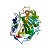
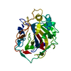
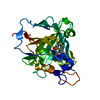
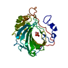
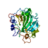
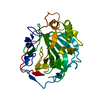
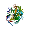
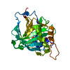
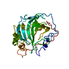
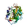
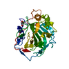
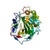
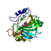
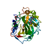
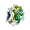
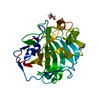
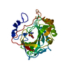
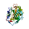
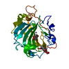
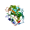
| Title | Human carbonic ahydrase II in complex with a benzenesulfonamide inhibitor | ||||||
 Components Components | Carbonic anhydrase 2 | ||||||
 Keywords Keywords | LYASE/LYASE INHIBITOR / lyase / benzenesulfonamide inhibitor / zinc metalloenzyme / zinc coordination / LYASE-LYASE INHIBITOR complex | ||||||
| Function / homology |  Function and homology information Function and homology informationpositive regulation of dipeptide transmembrane transport / : / secretion / regulation of monoatomic anion transport / cyanamide hydratase / cyanamide hydratase activity / arylesterase activity / regulation of chloride transport / Reversible hydration of carbon dioxide / positive regulation of synaptic transmission, GABAergic ...positive regulation of dipeptide transmembrane transport / : / secretion / regulation of monoatomic anion transport / cyanamide hydratase / cyanamide hydratase activity / arylesterase activity / regulation of chloride transport / Reversible hydration of carbon dioxide / positive regulation of synaptic transmission, GABAergic / morphogenesis of an epithelium / angiotensin-activated signaling pathway / Developmental Lineage of Pancreatic Ductal Cells / carbonic anhydrase / regulation of intracellular pH / carbonate dehydratase activity / carbon dioxide transport / neuron cellular homeostasis / Erythrocytes take up oxygen and release carbon dioxide / Erythrocytes take up carbon dioxide and release oxygen / apical part of cell / myelin sheath / extracellular exosome / zinc ion binding / plasma membrane / cytosol / cytoplasm Similarity search - Function | ||||||
| Biological species |  Homo sapiens (human) Homo sapiens (human) | ||||||
| Method |  X-RAY DIFFRACTION / MOLECULAR REPLACEMENT / Resolution: 1.498 Å X-RAY DIFFRACTION / MOLECULAR REPLACEMENT / Resolution: 1.498 Å | ||||||
 Authors Authors | Avvaru, B.S. / Wagner, J. / Robbins, A.H. / Mckenna, R. | ||||||
 Citation Citation | Journal: Chem.Commun.(Camb.) / Year: 2010 Title: Selective hydrophobic pocket binding observed within the carbonic anhydrase II active site accommodate different 4-substituted-ureido-benzenesulfonamides and correlate to inhibitor potency. Authors: Pacchiano, F. / Aggarwal, M. / Avvaru, B.S. / Robbins, A.H. / Scozzafava, A. / McKenna, R. / Supuran, C.T. | ||||||
| History |
|
- Structure visualization
| Structure viewer | Molecule: MolmilJmol/JSmol |
|---|
- Downloads & links
-Download
| PDBx/mmCIF format | 3mzc.cif.gz | 166.5 KB | Display | PDBx/mmCIF format |
|---|---|---|---|---|
| PDB format | pdb3mzc.ent.gz | 131.6 KB | Display | PDB format |
| PDBx/mmJSON format | 3mzc.json.gz | Tree view | PDBx/mmJSON format | |
| Others |  Other downloads Other downloads |
-Validation report
| Arichive directory | https://data.pdbj.org/pub/pdb/validation_reports/mz/3mzcftp://data.pdbj.org/pub/pdb/validation_reports/mz/3mzc | HTTPS FTP |
|---|
-Related structure data
| Related structure data |  3n0nC  3n2pC  3n3jC  3n4bC 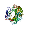 2iliS C: citing same article ( S: Starting model for refinement |
|---|---|
| Similar structure data |
-Links
 PDBj
PDBj

- Assembly
| Deposited unit | 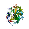
| ||||||||
|---|---|---|---|---|---|---|---|---|---|
| 1 |
| ||||||||
| Unit cell |
|
-Components
| #1: Protein | Mass: 29289.062 Da / Num. of mol.: 1 / Fragment: human carbonic anhydrase II Source method: isolated from a genetically manipulated source Source: (gene. exp.) Homo sapiens (human) / Gene: CAH2_HUMAN / Production host:  |
|---|---|
| #2: Chemical | ChemComp-ZN /   Mass: 65.409 Da / Num. of mol.: 1 / Source method: obtained synthetically / Formula: Zn Mass: 65.409 Da / Num. of mol.: 1 / Source method: obtained synthetically / Formula: Zn |
| #3: Chemical | ChemComp-S6I /   Mass: 283.347 Da / Num. of mol.: 1 / Source method: obtained synthetically / Formula: C12H17N3O3S Mass: 283.347 Da / Num. of mol.: 1 / Source method: obtained synthetically / Formula: C12H17N3O3S |
| #4: Chemical | ChemComp-GOL /   Mass: 92.094 Da / Num. of mol.: 1 / Fragment: sulfonamide inhibitor / Source method: obtained synthetically / Formula: C3H8O3 Mass: 92.094 Da / Num. of mol.: 1 / Fragment: sulfonamide inhibitor / Source method: obtained synthetically / Formula: C3H8O3 |
| #5: Water | ChemComp-HOH /  Mass: 18.015 Da / Num. of mol.: 306 / Source method: isolated from a natural source / Formula: H2O Mass: 18.015 Da / Num. of mol.: 306 / Source method: isolated from a natural source / Formula: H2O |
-Experimental details
-Experiment
| Experiment | Method: X-RAY DIFFRACTION / Number of used crystals: 1 |
|---|
- Sample preparation
| Crystal | Density Matthews: 2.1 Å3/Da / Density % sol: 41.38 % |
|---|---|
| Crystal grow | Temperature: 293 K / Method: vapor diffusion, hanging drop / pH: 7.8 Details: 1.2 M sodium citrate, 50 mM tris-HCL, 5uL + 5uL droplet, pH 7.8, VAPOR DIFFUSION, HANGING DROP, temperature 293K |
-Data collection
| Diffraction | Mean temperature: 100 K |
|---|---|
| Diffraction source | Source: ROTATING ANODE / Type: RIGAKU RUH3R / Wavelength: 1.5418 Å |
| Detector | Type: RIGAKU RAXIS IV / Detector: IMAGE PLATE / Date: Apr 12, 2010 / Details: mirrors |
| Radiation | Monochromator: Rigaku Optimax / Protocol: SINGLE WAVELENGTH / Monochromatic (M) / Laue (L): M / Scattering type: x-ray |
| Radiation wavelength | Wavelength: 1.5418 Å / Relative weight: 1 |
| Reflection | Resolution: 1.5→19.86 Å / Num. all: 35773 / Num. obs: 35690 / % possible obs: 91.4 % / Observed criterion σ(F): 0 / Observed criterion σ(I): 0 / Redundancy: 3.8 % / Biso Wilson estimate: 26.2 Å2 / Rmerge(I) obs: 0.041 / Rsym value: 0.041 / Χ2: 1.024 / Net I/σ(I): 24.4 |
| Reflection shell | Resolution: 1.5→1.55 Å / Redundancy: 3.9 % / Mean I/σ(I) obs: 4.9 / Num. unique all: 3407 / Rsym value: 0.253 / Χ2: 1.096 / % possible all: 87.9 |
- Processing
| Software |
| |||||||||||||||||||||||||||||||||||||||||||||||||||||||||||||||||||||||||||||||||||||||||||
|---|---|---|---|---|---|---|---|---|---|---|---|---|---|---|---|---|---|---|---|---|---|---|---|---|---|---|---|---|---|---|---|---|---|---|---|---|---|---|---|---|---|---|---|---|---|---|---|---|---|---|---|---|---|---|---|---|---|---|---|---|---|---|---|---|---|---|---|---|---|---|---|---|---|---|---|---|---|---|---|---|---|---|---|---|---|---|---|---|---|---|---|---|
| Refinement | Method to determine structure: MOLECULAR REPLACEMENT Starting model: PDB entry 2ILI Resolution: 1.498→19.868 Å / Occupancy max: 1 / Occupancy min: 0.38 / SU ML: 0.21 / Cross valid method: THROUGHOUT / σ(F): 0.07 / σ(I): 0 / Stereochemistry target values: ML
| |||||||||||||||||||||||||||||||||||||||||||||||||||||||||||||||||||||||||||||||||||||||||||
| Solvent computation | Shrinkage radii: 0.9 Å / VDW probe radii: 1.11 Å / Solvent model: FLAT BULK SOLVENT MODEL / Bsol: 48.18 Å2 / ksol: 0.483 e/Å3 | |||||||||||||||||||||||||||||||||||||||||||||||||||||||||||||||||||||||||||||||||||||||||||
| Displacement parameters | Biso max: 78.83 Å2 / Biso mean: 18.542 Å2 / Biso min: 6.56 Å2
| |||||||||||||||||||||||||||||||||||||||||||||||||||||||||||||||||||||||||||||||||||||||||||
| Refinement step | Cycle: LAST / Resolution: 1.498→19.868 Å
| |||||||||||||||||||||||||||||||||||||||||||||||||||||||||||||||||||||||||||||||||||||||||||
| Refine LS restraints |
| |||||||||||||||||||||||||||||||||||||||||||||||||||||||||||||||||||||||||||||||||||||||||||
| LS refinement shell | Refine-ID: X-RAY DIFFRACTION / Total num. of bins used: 12
| |||||||||||||||||||||||||||||||||||||||||||||||||||||||||||||||||||||||||||||||||||||||||||
| Refinement TLS params. | Method: refined / Refine-ID: X-RAY DIFFRACTION
| |||||||||||||||||||||||||||||||||||||||||||||||||||||||||||||||||||||||||||||||||||||||||||
| Refinement TLS group |
|
