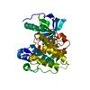
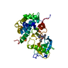
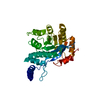
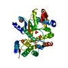
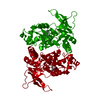
[English] 日本語
Yorodumi
- PDB-4jwx: GluN2A ligand-binding core in complex with propyl-NHP5G -

+
Open data


ID or keywords:

Loading...

-
Basic information

Entry
Database: PDB / ID: 4jwx
TitleGluN2A ligand-binding core in complex with propyl-NHP5G
ComponentsGluN2A
KeywordsUNKNOWN FUNCTION / bilobed structure
Function / homology
Function and homology information


neurotransmitter receptor transport, plasma membrane to endosome / regulation of response to alcohol / response to ammonium ion / receptor recycling / directional locomotion / response to environmental enrichment / auditory behavior / Assembly and cell surface presentation of NMDA receptors / serotonin metabolic process / response to hydrogen sulfide ...neurotransmitter receptor transport, plasma membrane to endosome / regulation of response to alcohol / response to ammonium ion / receptor recycling / directional locomotion / response to environmental enrichment / auditory behavior / Assembly and cell surface presentation of NMDA receptors / serotonin metabolic process / response to hydrogen sulfide / response to other organism / positive regulation of inhibitory postsynaptic potential / protein localization to postsynaptic membrane / cellular response to magnesium ion / regulation of ARF protein signal transduction / response to methylmercury / conditioned place preference / locomotion / dendritic spine organization / response to carbohydrate / regulation of NMDA receptor activity / sleep / cellular response to dsRNA / cellular response to lipid / Synaptic adhesion-like molecules / regulation of monoatomic cation transmembrane transport / NMDA glutamate receptor activity / voltage-gated monoatomic cation channel activity / response to manganese ion / NMDA selective glutamate receptor complex / cellular response to zinc ion / calcium ion transmembrane import into cytosol / glutamate receptor signaling pathway / glutamate binding / spinal cord development / parallel fiber to Purkinje cell synapse / regulation of postsynaptic membrane potential / response to amine / startle response / dopamine metabolic process / monoatomic cation transmembrane transport / response to lithium ion / modulation of excitatory postsynaptic potential / regulation of neuronal synaptic plasticity / action potential / cellular response to glycine / positive regulation of excitatory postsynaptic potential / response to light stimulus / positive regulation of protein targeting to membrane / Unblocking of NMDA receptors, glutamate binding and activation / cellular response to manganese ion / postsynaptic density, intracellular component / glutamate receptor binding / neuron development / multicellular organismal response to stress / monoatomic cation channel activity / glutamate-gated receptor activity / response to fungicide / cell adhesion molecule binding / glutamate-gated calcium ion channel activity / neurogenesis / ligand-gated monoatomic ion channel activity involved in regulation of presynaptic membrane potential / dendrite membrane / sensory perception of pain / sodium ion transmembrane transport / response to amphetamine / ionotropic glutamate receptor signaling pathway / cytoplasmic vesicle membrane / positive regulation of synaptic transmission, glutamatergic / protein tyrosine kinase binding / hippocampus development / learning / regulation of membrane potential / excitatory postsynaptic potential / transmitter-gated monoatomic ion channel activity involved in regulation of postsynaptic membrane potential / synaptic transmission, glutamatergic / cellular response to amino acid stimulus / response to nicotine / response to cocaine / regulation of long-term neuronal synaptic plasticity / synaptic membrane / modulation of chemical synaptic transmission / response to lead ion / protein catabolic process / postsynaptic density membrane / terminal bouton / : / visual learning / cellular response to growth factor stimulus / negative regulation of protein catabolic process / cerebral cortex development / calcium ion transmembrane transport / regulation of synaptic plasticity / calcium channel activity / memory / response to wounding / long-term synaptic potentiation / response to calcium ion / calcium ion transport / calcium-dependent protein binding
Similarity search - Function
Glutamate [NMDA] receptor, epsilon subunit, C-terminal / N-methyl D-aspartate receptor 2B3 C-terminus / Ionotropic glutamate receptor, metazoa / Ligated ion channel L-glutamate- and glycine-binding site / Ionotropic glutamate receptor, L-glutamate and glycine-binding domain / Ligated ion channel L-glutamate- and glycine-binding site / Ligand-gated ion channel / : / Ionotropic glutamate receptor / Eukaryotic homologues of bacterial periplasmic substrate binding proteins. ...Glutamate [NMDA] receptor, epsilon subunit, C-terminal / N-methyl D-aspartate receptor 2B3 C-terminus / Ionotropic glutamate receptor, metazoa / Ligated ion channel L-glutamate- and glycine-binding site / Ionotropic glutamate receptor, L-glutamate and glycine-binding domain / Ligated ion channel L-glutamate- and glycine-binding site / Ligand-gated ion channel / : / Ionotropic glutamate receptor / Eukaryotic homologues of bacterial periplasmic substrate binding proteins. / Receptor, ligand binding region / Receptor family ligand binding region / Periplasmic binding protein-like II / Periplasmic binding protein-like I / D-Maltodextrin-Binding Protein; domain 2 / 3-Layer(aba) Sandwich / Alpha Beta
Similarity search - Domain/homology
Chem-1N4 / Glutamate receptor ionotropic, NMDA 2A
Similarity search - Component
Biological speciesRattus norvegicus (Norway rat)
MethodX-RAY DIFFRACTION / SYNCHROTRON / MOLECULAR REPLACEMENT / Resolution: 1.5 Å
AuthorsHansen, K.B. / Tajima, N. / Risgaard, R. / Perszyk, R.E. / Jorgensen, L. / Vance, K.M. / Ogden, K.K. / Clausen, R.P. / Furukawa, H. / Traynelis, S.F.
CitationJournal: Mol.Pharmacol. / Year: 2013
Title: Structural determinants of agonist efficacy at the glutamate binding site of N-methyl-d-aspartate receptors.
Authors: Hansen, K.B. / Tajima, N. / Risgaard, R. / Perszyk, R.E. / Jorgensen, L. / Vance, K.M. / Ogden, K.K. / Clausen, R.P. / Furukawa, H. / Traynelis, S.F.
History
DepositionMar 27, 2013Deposition site: RCSB / Processing site: RCSB
Revision 1.0May 29, 2013Provider: repository / Type: Initial release
Revision 1.1Jul 17, 2013Group: Database references
Revision 1.2Sep 20, 2023Group: Data collection / Database references ...Data collection / Database references / Derived calculations / Refinement description
Category: chem_comp_atom / chem_comp_bond ...chem_comp_atom / chem_comp_bond / database_2 / pdbx_initial_refinement_model / struct_site
Item: _database_2.pdbx_DOI / _database_2.pdbx_database_accession ..._database_2.pdbx_DOI / _database_2.pdbx_database_accession / _struct_site.pdbx_auth_asym_id / _struct_site.pdbx_auth_comp_id / _struct_site.pdbx_auth_seq_id
Revision 1.3Oct 9, 2024Group: Structure summary / Category: pdbx_entry_details / pdbx_modification_feature

-
Structure visualization

Structure viewerMolecule:
MolmilJmol/JSmol

Downloads & links

-
Assembly

Deposited unit
A: GluN2A
hetero molecules


Theoretical massNumber of molelcules
Total (without water)31,6242
Polymers31,4251
Non-polymers1991
Water8,395466
1


  • Idetical with deposited unit
  • defined by author
TypeNameSymmetry operationNumber
identity operation1_555x,y,z1
2
A: GluN2A
hetero molecules

A: GluN2A
hetero molecules


Theoretical massNumber of molelcules
Total (without water)63,2484
Polymers62,8502
Non-polymers3982
Water362
TypeNameSymmetry operationNumber
identity operation1_555x,y,z1
crystal symmetry operation8_665-y+1,-x+1,-z+1/21
Buried area3030 Å2
ΔGint-15 kcal/mol
Surface area26700 Å2
MethodPISA
Unit cell
Length a, b, c (Å)52.031, 52.031, 198.823
Angle α, β, γ (deg.)90.00, 90.00, 90.00
Int Tables number92
Space group name H-MP41212

-
Components

#1: Protein GluN2A


Mass: 31425.018 Da / Num. of mol.: 1 / Fragment: ligand binding domain
Source method: isolated from a genetically manipulated source
Source: (gene. exp.) Rattus norvegicus (Norway rat) / Plasmid: pET22 / Production host: Escherichia coli (E. coli) / Strain (production host): Origami B (DE3) / References: UniProt: Q00959*PLUS
#2: Chemical ChemComp-1N4 / (2R)-amino(1-hydroxy-4-propyl-1H-pyrazol-5-yl)ethanoic acid


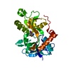
Mass: 199.207 Da / Num. of mol.: 1 / Source method: obtained synthetically / Formula: C8H13N3O3
#3: Water ChemComp-HOH / water


Mass: 18.015 Da / Num. of mol.: 466 / Source method: isolated from a natural source / Formula: H2O
Has protein modificationY

-
Experimental details

-
Experiment

ExperimentMethod: X-RAY DIFFRACTION / Number of used crystals: 1

-
Sample preparation

CrystalDensity Matthews: 2.14 Å3/Da / Density % sol: 42.55 %
Crystal growTemperature: 291 K / Method: vapor diffusion, hanging drop / pH: 7
Details: 100 mM HEPES-NaOH, 10-18% PEG8000, 100 mM calcium acatate, pH 7.0, VAPOR DIFFUSION, HANGING DROP, temperature 291K

-
Data collection

DiffractionMean temperature: 100 K
Diffraction sourceSource: SYNCHROTRON / Site: NSLS / Beamline: X25 / Wavelength: 1.1 Å
RadiationProtocol: SINGLE WAVELENGTH / Monochromatic (M) / Laue (L): M / Scattering type: x-ray
Radiation wavelengthWavelength: 1.1 Å / Relative weight: 1
ReflectionResolution: 1.5→50 Å / Num. all: 45212 / Num. obs: 44489 / % possible obs: 98.4 % / Observed criterion σ(F): 2 / Observed criterion σ(I): 2 / Redundancy: 12.6 %

-
Processing

Software
NameVersionClassification
HKL-2000data collection
PHASERphasing
PHENIX(phenix.refine: 1.8.1_1168)refinement
HKL-2000data reduction
HKL-2000data scaling
RefinementMethod to determine structure: MOLECULAR REPLACEMENT
Starting model: PDB entry 1FTJ
Resolution: 1.5→19.184 Å / SU ML: 0.1 / σ(F): 1.34 / σ(I): 2 / Phase error: 17.4 / Stereochemistry target values: ML
RfactorNum. reflection% reflectionSelection details
Rfree0.1946 1895 5.09 %random
Rwork0.1597 ---
obs0.1615 37218 99.95 %-
all-37237 --
Solvent computationShrinkage radii: 0.9 Å / VDW probe radii: 1.11 Å / Solvent model: FLAT BULK SOLVENT MODEL
Refinement stepCycle: LAST / Resolution: 1.5→19.184 Å
ProteinNucleic acidLigandSolventTotal
Num. atoms2204 0 14 466 2684
Refine LS restraints
Refine-IDTypeDev idealNumber
X-RAY DIFFRACTIONf_bond_d0.0072454
X-RAY DIFFRACTIONf_angle_d1.1453359
X-RAY DIFFRACTIONf_dihedral_angle_d13.562950
X-RAY DIFFRACTIONf_chiral_restr0.08384
X-RAY DIFFRACTIONf_plane_restr0.005431
LS refinement shell
Resolution (Å)Rfactor RfreeNum. reflection RfreeRfactor RworkNum. reflection RworkRefine-ID% reflection obs (%)
1.5-1.640.20251340.17472444X-RAY DIFFRACTION100
1.64-1.68430.20691340.16842489X-RAY DIFFRACTION100
1.6843-1.73390.21881400.17272451X-RAY DIFFRACTION100
1.7339-1.78980.24181300.17332485X-RAY DIFFRACTION100
1.7898-1.85370.19821200.17262479X-RAY DIFFRACTION100
1.8537-1.92790.17831340.1652493X-RAY DIFFRACTION100
1.9279-2.01550.19781350.16352475X-RAY DIFFRACTION100
2.0155-2.12160.20061300.1582495X-RAY DIFFRACTION100
2.1216-2.25440.17171300.15992524X-RAY DIFFRACTION100
2.2544-2.42810.22531540.16472494X-RAY DIFFRACTION100
2.4281-2.67190.20931400.16432533X-RAY DIFFRACTION100
2.6719-3.05720.21081440.16832578X-RAY DIFFRACTION100
3.0572-3.84660.18161220.14442606X-RAY DIFFRACTION100
3.8466-19.18580.16511480.15012777X-RAY DIFFRACTION100

+
About Yorodumi

-
News

-
Feb 9, 2022. New format data for meta-information of EMDB entries

New format data for meta-information of EMDB entries

  • Version 3 of the EMDB header file is now the official format.
  • The previous official version 1.9 will be removed from the archive.

Related info.:EMDB header

External links:wwPDB to switch to version 3 of the EMDB data model

-
Aug 12, 2020. Covid-19 info

Covid-19 info

URL: https://pdbj.org/emnavi/covid19.php

New page: Covid-19 featured information page in EM Navigator.

Related info.:Covid-19 info / Mar 5, 2020. Novel coronavirus structure data

+
Mar 5, 2020. Novel coronavirus structure data

Novel coronavirus structure data

Related info.:Yorodumi Speices / Aug 12, 2020. Covid-19 info

External links:COVID-19 featured content - PDBj / Molecule of the Month (242):Coronavirus Proteases

+
Jan 31, 2019. EMDB accession codes are about to change! (news from PDBe EMDB page)

EMDB accession codes are about to change! (news from PDBe EMDB page)

  • The allocation of 4 digits for EMDB accession codes will soon come to an end. Whilst these codes will remain in use, new EMDB accession codes will include an additional digit and will expand incrementally as the available range of codes is exhausted. The current 4-digit format prefixed with “EMD-” (i.e. EMD-XXXX) will advance to a 5-digit format (i.e. EMD-XXXXX), and so on. It is currently estimated that the 4-digit codes will be depleted around Spring 2019, at which point the 5-digit format will come into force.
  • The EM Navigator/Yorodumi systems omit the EMD- prefix.

Related info.:Q: What is EMD? / ID/Accession-code notation in Yorodumi/EM Navigator

External links:EMDB Accession Codes are Changing Soon! / Contact to PDBj

+
Jul 12, 2017. Major update of PDB

Major update of PDB

  • wwPDB released updated PDB data conforming to the new PDBx/mmCIF dictionary.
  • This is a major update changing the version number from 4 to 5, and with Remediation, in which all the entries are updated.
  • In this update, many items about electron microscopy experimental information are reorganized (e.g. em_software).
  • Now, EM Navigator and Yorodumi are based on the updated data.

External links:wwPDB Remediation / Enriched Model Files Conforming to OneDep Data Standards Now Available in the PDB FTP Archive

-
Yorodumi

Thousand views of thousand structures

  • Yorodumi is a browser for structure data from EMDB, PDB, SASBDB, etc.
  • This page is also the successor to EM Navigator detail page, and also detail information page/front-end page for Omokage search.
  • The word "yorodu" (or yorozu) is an old Japanese word meaning "ten thousand". "mi" (miru) is to see.

Related info.:EMDB / PDB / SASBDB / Comparison of 3 databanks / Yorodumi Search / Aug 31, 2016. New EM Navigator & Yorodumi / Yorodumi Papers / Jmol/JSmol / Function and homology information / Changes in new EM Navigator and Yorodumi

Read more