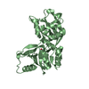
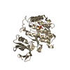
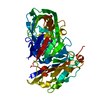
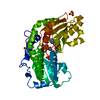
Entry Database : PDB / ID : 2a5sTitle Crystal Structure Of The NR2A Ligand Binding Core In Complex With Glutamate N-methyl-D-aspartate receptor NMDAR2A subunit Keywords / / Function / homology Function Domain/homology Component
/ / / / / / / / / / / / / / / / / / / / / / / / / / / / / / / / / / / / / / / / / / / / / / / / / / / / / / / / / / / / / / / / / / / / / / / / / / / / / / / / / / / / / / / / / / / / / / / / / / / / / / / / / / / / / / / / / / / / / / / / / / / / / / / / / / / / / Biological species Rattus norvegicus (Norway rat)Method / / / Resolution : 1.7 Å Authors Furukawa, H. / Singh, S.K. / Mancusso, R. / Gouaux, E. Journal : Nature / Year : 2005Title : Subunit arrangement and function in NMDA receptorsAuthors : Furukawa, H. / Singh, S.K. / Mancusso, R. / Gouaux, E. History Deposition Jun 30, 2005 Deposition site / Processing site Revision 1.0 Nov 15, 2005 Provider / Type Revision 1.1 Apr 30, 2008 Group Revision 1.2 Jul 13, 2011 Group Revision 1.3 Jul 26, 2017 Group / Source and taxonomy / Category / softwareRevision 1.4 Oct 30, 2024 Group Data collection / Database references ... Data collection / Database references / Derived calculations / Structure summary Category chem_comp_atom / chem_comp_bond ... chem_comp_atom / chem_comp_bond / database_2 / pdbx_entry_details / pdbx_modification_feature / struct_site Item _database_2.pdbx_DOI / _database_2.pdbx_database_accession ... _database_2.pdbx_DOI / _database_2.pdbx_database_accession / _struct_site.pdbx_auth_asym_id / _struct_site.pdbx_auth_comp_id / _struct_site.pdbx_auth_seq_id
Show all Show less
 Movie
Movie Controller

 Yorodumi
 Open data
 Basic information
 Components
 Keywords
 Function and homology information
 X-RAY DIFFRACTION /
  Authors
 Citation
 Structure visualization
 Downloads & links
 Other downloads


 PDBj
PDBj


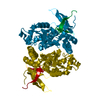
 Assembly


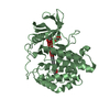
 Type: L-peptide linking / Mass: 147.129 Da / Num. of mol.: 1 / Source method: obtained synthetically / Formula: C5H9NO4
 Mass: 18.015 Da / Num. of mol.: 316 / Source method: isolated from a natural source / Formula: H2O
 Sample preparation
 / Beamline: X4A / Wavelength: 0.9793 Å
 Processing