stress response to copper ion / sigma factor antagonist activity / cellular response to unfolded protein / protein unfolding / heat shock protein binding / protein folding chaperone / inclusion body / ATP-dependent protein folding chaperone / ADP binding / : ...stress response to copper ion / sigma factor antagonist activity / cellular response to unfolded protein / protein unfolding / heat shock protein binding / protein folding chaperone / inclusion body / ATP-dependent protein folding chaperone / ADP binding / : / response to heat / protein refolding / protein-folding chaperone binding / protein folding / protein-containing complex assembly / DNA replication / defense response to bacterium / ATP hydrolysis activity / protein-containing complex / extracellular region / zinc ion binding / ATP binding / membrane / plasma membrane / cytosol / cytoplasm Similarity search - Function
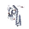
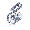
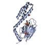
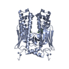
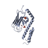
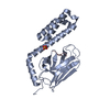
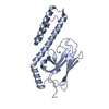
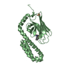
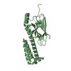
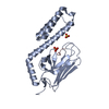
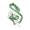
Cathelicidins signature 1. / Cathelicidin, conserved site / Cathelicidins signature 2. / Cathelicidin-like / Cathelicidin / Substrate Binding Domain Of Dnak; Chain:A; Domain 2 - #10 / Substrate Binding Domain Of DNAk; Chain A, domain 1 / Chaperone DnaK / Cystatin superfamily / Substrate Binding Domain Of DNAk; Chain A, domain 1 ...Cathelicidins signature 1. / Cathelicidin, conserved site / Cathelicidins signature 2. / Cathelicidin-like / Cathelicidin / Substrate Binding Domain Of Dnak; Chain:A; Domain 2 - #10 / Substrate Binding Domain Of DNAk; Chain A, domain 1 / Chaperone DnaK / Cystatin superfamily / Substrate Binding Domain Of DNAk; Chain A, domain 1 / Substrate Binding Domain Of Dnak; Chain:A; Domain 2 / Heat shock hsp70 proteins family signature 2. / Heat shock hsp70 proteins family signature 1. / Heat shock hsp70 proteins family signature 3. / Heat shock protein 70, conserved site / Heat shock protein 70kD, peptide-binding domain superfamily / Heat shock protein 70kD, C-terminal domain superfamily / Heat shock protein 70 family / Hsp70 protein / ATPase, nucleotide binding domain / Up-down Bundle / Sandwich / Mainly Beta / Mainly Alpha Similarity search - Domain/homology
In the structure databanks used in Yorodumi, some data are registered as the other names, "COVID-19 virus" and "2019-nCoV". Here are the details of the virus and the list of structure data.
Jan 31, 2019. EMDB accession codes are about to change! (news from PDBe EMDB page)
EMDB accession codes are about to change! (news from PDBe EMDB page)
The allocation of 4 digits for EMDB accession codes will soon come to an end. Whilst these codes will remain in use, new EMDB accession codes will include an additional digit and will expand incrementally as the available range of codes is exhausted. The current 4-digit format prefixed with “EMD-” (i.e. EMD-XXXX) will advance to a 5-digit format (i.e. EMD-XXXXX), and so on. It is currently estimated that the 4-digit codes will be depleted around Spring 2019, at which point the 5-digit format will come into force.
The EM Navigator/Yorodumi systems omit the EMD- prefix.
Related info.:Q: What is EMD? / ID/Accession-code notation in Yorodumi/EM Navigator
Yorodumi is a browser for structure data from EMDB, PDB, SASBDB, etc.
This page is also the successor to EM Navigator detail page, and also detail information page/front-end page for Omokage search.
The word "yorodu" (or yorozu) is an old Japanese word meaning "ten thousand". "mi" (miru) is to see.
Related info.:EMDB / PDB / SASBDB / Comparison of 3 databanks / Yorodumi Search / Aug 31, 2016. New EM Navigator & Yorodumi / Yorodumi Papers / Jmol/JSmol / Function and homology information / Changes in new EM Navigator and Yorodumi
 Movie
Movie Controller

 Yorodumi
 Open data
 Basic information
 Components
 Keywords
 Function and homology information

 X-RAY DIFFRACTION /
  Authors
 Citation
 Structure visualization
 Downloads & links
 Other downloads


 PDBj
PDBj

 Assembly


 Mass: 96.063 Da / Num. of mol.: 3 / Source method: obtained synthetically / Formula: SO4
 Mass: 18.015 Da / Num. of mol.: 298 / Source method: isolated from a natural source / Formula: H2O
 Sample preparation
 / Beamline: 14.1 / Wavelength: 0.918 Å
 Processing