 Movie
Movie Controller

[English] 日本語
 Yorodumi
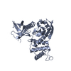
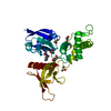
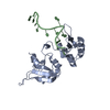
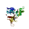
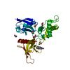
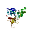
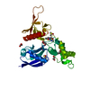
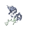
- PDB-4jw2: Selection of specific protein binders for pre-defined targets fro... -
+ Open data

- Basic information
| Entry | Database: PDB / ID: 4jw2 | ||||||
|---|---|---|---|---|---|---|---|
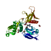
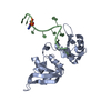
| Title | Selection of specific protein binders for pre-defined targets from an optimized library of artificial helicoidal repeat proteins (alphaRep) | ||||||
 Components Components |
| ||||||
 Keywords Keywords | DE NOVO PROTEIN/protein binding / alpha-helical proteins / Proteins engineered to bind to various partners / DE NOVO PROTEIN-protein binding complex | ||||||
| Function / homology | Leucine-rich Repeat Variant / Leucine-rich Repeat Variant / Alpha Horseshoe / Mainly Alpha Function and homology information Function and homology information | ||||||
| Biological species | synthetic construct (others) | ||||||
| Method |  X-RAY DIFFRACTION / SYNCHROTRON / MOLECULAR REPLACEMENT / Resolution: 1.9 Å X-RAY DIFFRACTION / SYNCHROTRON / MOLECULAR REPLACEMENT / Resolution: 1.9 Å | ||||||
 Authors Authors | Guellouz, A. / Valerio-Lepiniec, M. / Urvoas, A. / Chevrel, A. / Graille, M. / Fourati-Kammoun, Z. / Desmadril, M. / van Tilbeurgh, H. / Minard, P. | ||||||
 Citation Citation | Journal: Plos One / Year: 2013 Title: Selection of Specific Protein Binders for Pre-Defined Targets from an Optimized Library of Artificial Helicoidal Repeat Proteins (alphaRep). Authors: Guellouz, A. / Valerio-Lepiniec, M. / Urvoas, A. / Chevrel, A. / Graille, M. / Fourati-Kammoun, Z. / Desmadril, M. / van Tilbeurgh, H. / Minard, P. | ||||||
| History |
|
- Structure visualization
| Structure viewer | Molecule: MolmilJmol/JSmol |
|---|
- Downloads & links
-Download
| PDBx/mmCIF format | 4jw2.cif.gz | 66.3 KB | Display | PDBx/mmCIF format |
|---|---|---|---|---|
| PDB format | pdb4jw2.ent.gz | 48.8 KB | Display | PDB format |
| PDBx/mmJSON format | 4jw2.json.gz | Tree view | PDBx/mmJSON format | |
| Others |  Other downloads Other downloads |
-Validation report
| Arichive directory | https://data.pdbj.org/pub/pdb/validation_reports/jw/4jw2ftp://data.pdbj.org/pub/pdb/validation_reports/jw/4jw2 | HTTPS FTP |
|---|
-Related structure data
| Related structure data |  4jw3C  3ltmS S: Starting model for refinement C: citing same article ( |
|---|---|
| Similar structure data |
-Links
 PDBj
PDBj- Assembly
| Deposited unit | 
| ||||||||
|---|---|---|---|---|---|---|---|---|---|
| 1 |
| ||||||||
| Unit cell |
|
-Components
| #1: Protein | Mass: 11947.639 Da / Num. of mol.: 1 Source method: isolated from a genetically manipulated source Source: (gene. exp.) synthetic construct (others) / Plasmid: pQE81L / Production host:  | ||
|---|---|---|---|
| #2: Protein | Mass: 22224.102 Da / Num. of mol.: 1 Source method: isolated from a genetically manipulated source Source: (gene. exp.) synthetic construct (others) / Plasmid: pQE81L / Production host: | ||
| #3: Chemical |   Mass: 62.068 Da / Num. of mol.: 3 / Source method: obtained synthetically / Formula: C2H6O2 Mass: 62.068 Da / Num. of mol.: 3 / Source method: obtained synthetically / Formula: C2H6O2#4: Water | ChemComp-HOH / |  Mass: 18.015 Da / Num. of mol.: 146 / Source method: isolated from a natural source / Formula: H2O Mass: 18.015 Da / Num. of mol.: 146 / Source method: isolated from a natural source / Formula: H2O |
-Experimental details
-Experiment
| Experiment | Method: X-RAY DIFFRACTION / Number of used crystals: 1 |
|---|
- Sample preparation
| Crystal | Density Matthews: 2.2 Å3/Da / Density % sol: 44.01 % |
|---|---|
| Crystal grow | Temperature: 293 K / Method: vapor diffusion, sitting drop / pH: 4.6 Details: 8% (w/v) PEG 2,000 MME, 100 mM Na acetate pH 4.6, VAPOR DIFFUSION, SITTING DROP, temperature 293K |
-Data collection
| Diffraction | Mean temperature: 100 K |
|---|---|
| Diffraction source | Source: SYNCHROTRON / Site: SOLEIL  / Beamline: PROXIMA 1 / Wavelength: 0.97918 Å / Beamline: PROXIMA 1 / Wavelength: 0.97918 Å |
| Detector | Type: PSI PILATUS 6M / Detector: PIXEL / Date: Feb 29, 2012 |
| Radiation | Monochromator: Si 111 CHANNEL / Protocol: SINGLE WAVELENGTH / Monochromatic (M) / Laue (L): M / Scattering type: x-ray |
| Radiation wavelength | Wavelength: 0.97918 Å / Relative weight: 1 |
| Reflection | Resolution: 1.9→30 Å / Num. all: 24720 / Num. obs: 24498 / % possible obs: 99.1 % / Observed criterion σ(F): 2 / Observed criterion σ(I): 2 / Redundancy: 5.5 % / Biso Wilson estimate: 36.05 Å2 / Rsym value: 0.048 / Net I/σ(I): 17.6 |
| Reflection shell | Resolution: 1.9→1.95 Å / Redundancy: 5.5 % / Mean I/σ(I) obs: 2 / Rsym value: 0.566 / % possible all: 98.9 |
- Processing
| Software |
| ||||||||||||||||||||||||||||||||||||||||||||||||||||||||||||||||||
|---|---|---|---|---|---|---|---|---|---|---|---|---|---|---|---|---|---|---|---|---|---|---|---|---|---|---|---|---|---|---|---|---|---|---|---|---|---|---|---|---|---|---|---|---|---|---|---|---|---|---|---|---|---|---|---|---|---|---|---|---|---|---|---|---|---|---|---|
| Refinement | Method to determine structure: MOLECULAR REPLACEMENT Starting model: 3LTM Resolution: 1.9→28.83 Å / Cor.coef. Fo:Fc: 0.9204 / Cor.coef. Fo:Fc free: 0.9132 / Cross valid method: THROUGHOUT / σ(F): 0 / Stereochemistry target values: Engh & Huber
| ||||||||||||||||||||||||||||||||||||||||||||||||||||||||||||||||||
| Displacement parameters | Biso mean: 48.27 Å2
| ||||||||||||||||||||||||||||||||||||||||||||||||||||||||||||||||||
| Refine analyze | Luzzati coordinate error obs: 0.261 Å | ||||||||||||||||||||||||||||||||||||||||||||||||||||||||||||||||||
| Refinement step | Cycle: LAST / Resolution: 1.9→28.83 Å
| ||||||||||||||||||||||||||||||||||||||||||||||||||||||||||||||||||
| Refine LS restraints |
| ||||||||||||||||||||||||||||||||||||||||||||||||||||||||||||||||||
| LS refinement shell | Resolution: 1.9→1.98 Å / Total num. of bins used: 12
|
