 Movie
Movie Controller

[English] 日本語
 Yorodumi
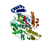
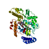
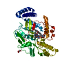
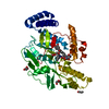
- PDB-3ltm: Structure of a new family of artificial alpha helicoidal repeat p... -
+ Open data

- Basic information
| Entry | Database: PDB / ID: 3ltm | ||||||
|---|---|---|---|---|---|---|---|
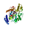
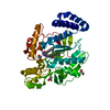
| Title | Structure of a new family of artificial alpha helicoidal repeat proteins (alpha-Rep) based on thermostable HEAT-like repeats | ||||||
 Components Components | Alpha-Rep4 | ||||||
 Keywords Keywords | PROTEIN BINDING / Protein engineering / HEAT-like repeat | ||||||
| Function / homology | Leucine-rich Repeat Variant / Leucine-rich Repeat Variant / Alpha Horseshoe / Mainly Alpha Function and homology information Function and homology information | ||||||
| Biological species | Synthetic (others) | ||||||
| Method |  X-RAY DIFFRACTION / SYNCHROTRON / MAD / Resolution: 2.15 Å X-RAY DIFFRACTION / SYNCHROTRON / MAD / Resolution: 2.15 Å | ||||||
 Authors Authors | Urvoas, A. / Guellouz, A. / Graille, M. / van Tilbeurgh, H. / Desmadril, M. / Minard, P. | ||||||
 Citation Citation | Journal: J.Mol.Biol. / Year: 2010 Title: Design, production and molecular structure of a new family of artificial alpha-helicoidal repeat proteins ( alpha Rep) based on thermostable HEAT-like repeats Authors: Urvoas, A. / Guellouz, A. / Valerio-Lepiniec, M. / Graille, M. / Durand, D. / Desravines, D.C. / van Tilbeurgh, H. / Desmadril, M. / Minard, P. | ||||||
| History |
|
- Structure visualization
| Structure viewer | Molecule: MolmilJmol/JSmol |
|---|
- Downloads & links
-Download
| PDBx/mmCIF format | 3ltm.cif.gz | 93.3 KB | Display | PDBx/mmCIF format |
|---|---|---|---|---|
| PDB format | pdb3ltm.ent.gz | 70.1 KB | Display | PDB format |
| PDBx/mmJSON format | 3ltm.json.gz | Tree view | PDBx/mmJSON format | |
| Others |  Other downloads Other downloads |
-Validation report
| Arichive directory | https://data.pdbj.org/pub/pdb/validation_reports/lt/3ltmftp://data.pdbj.org/pub/pdb/validation_reports/lt/3ltm | HTTPS FTP |
|---|
-Related structure data


-Links
 PDBj
PDBj- Assembly
| Deposited unit | 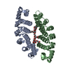
| ||||||||
|---|---|---|---|---|---|---|---|---|---|
| 1 |
| ||||||||
| Unit cell |
|
-Components
| #1: Protein | Mass: 23403.359 Da / Num. of mol.: 2 Source method: isolated from a genetically manipulated source Source: (gene. exp.) Synthetic (others) / Production host:  #2: Chemical | ChemComp-1PE / |   Mass: 238.278 Da / Num. of mol.: 1 / Source method: obtained synthetically / Formula: C10H22O6 / Comment: precipitant*YM Mass: 238.278 Da / Num. of mol.: 1 / Source method: obtained synthetically / Formula: C10H22O6 / Comment: precipitant*YM#3: Chemical | ChemComp-12P / | 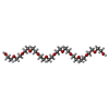  Mass: 546.646 Da / Num. of mol.: 1 / Source method: obtained synthetically / Formula: C24H50O13 / Comment: precipitant*YM Mass: 546.646 Da / Num. of mol.: 1 / Source method: obtained synthetically / Formula: C24H50O13 / Comment: precipitant*YM#4: Chemical |   Mass: 92.094 Da / Num. of mol.: 2 / Source method: obtained synthetically / Formula: C3H8O3 Mass: 92.094 Da / Num. of mol.: 2 / Source method: obtained synthetically / Formula: C3H8O3#5: Water | ChemComp-HOH / |  Mass: 18.015 Da / Num. of mol.: 331 / Source method: isolated from a natural source / Formula: H2O Mass: 18.015 Da / Num. of mol.: 331 / Source method: isolated from a natural source / Formula: H2O |
|---|
-Experimental details
-Experiment
| Experiment | Method: X-RAY DIFFRACTION / Number of used crystals: 1 |
|---|
- Sample preparation
| Crystal | Density Matthews: 2 Å3/Da / Density % sol: 38.38 % |
|---|---|
| Crystal grow | Temperature: 292 K / Method: vapor diffusion, sitting drop / pH: 8 Details: 18.5% PEG 1000, 6.5% glycerol, 100 mM tricine pH8, 230 mM NaCl, VAPOR DIFFUSION, SITTING DROP, temperature 292K |
-Data collection
| Diffraction | Mean temperature: 100 K | ||||||||||||
|---|---|---|---|---|---|---|---|---|---|---|---|---|---|
| Diffraction source | Source: SYNCHROTRON / Site: SOLEIL  / Beamline: PROXIMA 1 / Wavelength: 0.91971, 0.9795, 0.93 / Beamline: PROXIMA 1 / Wavelength: 0.91971, 0.9795, 0.93 | ||||||||||||
| Detector | Type: ADSC QUANTUM 315r / Detector: CCD / Date: Dec 12, 2008 | ||||||||||||
| Radiation | Monochromator: SAGITALLY FOCUSED Si(111) / Protocol: MAD / Monochromatic (M) / Laue (L): M / Scattering type: x-ray | ||||||||||||
| Radiation wavelength |
| ||||||||||||
| Reflection | Resolution: 2.15→30 Å / Num. obs: 20242 / % possible obs: 99.7 % / Observed criterion σ(F): 0 / Observed criterion σ(I): 0 / Redundancy: 3.7 % / Rsym value: 0.079 / Net I/σ(I): 14.4 | ||||||||||||
| Reflection shell | Resolution: 2.15→2.21 Å / Redundancy: 3.7 % / Mean I/σ(I) obs: 3.2 / Rsym value: 0.446 / % possible all: 99.5 |
- Processing
| Software |
| ||||||||||||||||||||||||||||||||||||||||||||||||||||||||
|---|---|---|---|---|---|---|---|---|---|---|---|---|---|---|---|---|---|---|---|---|---|---|---|---|---|---|---|---|---|---|---|---|---|---|---|---|---|---|---|---|---|---|---|---|---|---|---|---|---|---|---|---|---|---|---|---|---|
| Refinement | Method to determine structure: MAD / Resolution: 2.15→19.802 Å / Occupancy max: 1 / Occupancy min: 0.08 / FOM work R set: 0.807 / SU ML: 0.33 / Cross valid method: THROUGHOUT / σ(F): 1.99 / Phase error: 26.06 / Stereochemistry target values: ML
| ||||||||||||||||||||||||||||||||||||||||||||||||||||||||
| Solvent computation | Shrinkage radii: 0.9 Å / VDW probe radii: 1.11 Å / Solvent model: FLAT BULK SOLVENT MODEL / Bsol: 57.149 Å2 / ksol: 0.382 e/Å3 | ||||||||||||||||||||||||||||||||||||||||||||||||||||||||
| Displacement parameters | Biso max: 151.78 Å2 / Biso mean: 33.584 Å2 / Biso min: 5.1 Å2
| ||||||||||||||||||||||||||||||||||||||||||||||||||||||||
| Refinement step | Cycle: LAST / Resolution: 2.15→19.802 Å
| ||||||||||||||||||||||||||||||||||||||||||||||||||||||||
| Refine LS restraints |
| ||||||||||||||||||||||||||||||||||||||||||||||||||||||||
| LS refinement shell |
|
