 Movie
Movie Controller

[English] 日本語
 Yorodumi
- PDB-1u1o: Crystal Structure of UP1 Complexed With d(TTAGGGTTAG(DI)G); A Hum... -
+ Open data

- Basic information
| Entry | Database: PDB / ID: 1u1o | ||||||
|---|---|---|---|---|---|---|---|
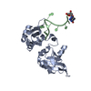
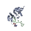
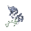
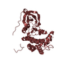
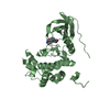
| Title | Crystal Structure of UP1 Complexed With d(TTAGGGTTAG(DI)G); A Human Telomeric Repeat Containing Inosine | ||||||
 Components Components |
| ||||||
 Keywords Keywords | TRANSPORT PROTEIN/DNA / protein-DNA complex / UP1 / human telomeric repeat / hTR / TR2-G(11)DI / RRM / RNA Recognition Motif / DI / inosine / hnRNP A1 / TRANSPORT PROTEIN-DNA COMPLEX | ||||||
| Function / homology |  Function and homology information Function and homology informationcellular response to sodium arsenite / SARS-CoV-1-host interactions / import into nucleus / telomeric repeat-containing RNA binding / alternative mRNA splicing, via spliceosome / pre-mRNA binding / G-rich strand telomeric DNA binding / nuclear export / RNA export from nucleus / FGFR2 alternative splicing ...cellular response to sodium arsenite / SARS-CoV-1-host interactions / import into nucleus / telomeric repeat-containing RNA binding / alternative mRNA splicing, via spliceosome / pre-mRNA binding / G-rich strand telomeric DNA binding / nuclear export / RNA export from nucleus / FGFR2 alternative splicing / miRNA binding / regulation of alternative mRNA splicing, via spliceosome / regulation of RNA splicing / negative regulation of telomere maintenance via telomerase / Processing of Capped Intron-Containing Pre-mRNA / SARS-CoV-1 modulates host translation machinery / mRNA transport / cellular response to glucose starvation / catalytic step 2 spliceosome / positive regulation of telomere maintenance via telomerase / mRNA Splicing - Major Pathway / spliceosomal complex / mRNA 3'-UTR binding / mRNA splicing, via spliceosome / single-stranded DNA binding / single-stranded RNA binding / ribonucleoprotein complex / protein domain specific binding / synapse / DNA binding / RNA binding / extracellular exosome / nucleoplasm / membrane / identical protein binding / nucleus / cytoplasm / cytosol Similarity search - Function | ||||||
| Biological species |  Homo sapiens (human) Homo sapiens (human) | ||||||
| Method |  X-RAY DIFFRACTION / MOLECULAR REPLACEMENT / Resolution: 2 Å X-RAY DIFFRACTION / MOLECULAR REPLACEMENT / Resolution: 2 Å | ||||||
 Authors Authors | Myers, J.C. / Shamoo, Y. | ||||||
 Citation Citation | Journal: J.Mol.Biol. / Year: 2004 Title: Human UP1 as a Model for Understanding Purine Recognition in the Family of Proteins Containing the RNA Recognition Motif (RRM). Authors: Myers, J.C. / Shamoo, Y. | ||||||
| History |
|
- Structure visualization
| Structure viewer | Molecule: MolmilJmol/JSmol |
|---|
- Downloads & links
-Download
| PDBx/mmCIF format | 1u1o.cif.gz | 57.9 KB | Display | PDBx/mmCIF format |
|---|---|---|---|---|
| PDB format | pdb1u1o.ent.gz | 40.1 KB | Display | PDB format |
| PDBx/mmJSON format | 1u1o.json.gz | Tree view | PDBx/mmJSON format | |
| Others |  Other downloads Other downloads |
-Validation report
| Arichive directory | https://data.pdbj.org/pub/pdb/validation_reports/u1/1u1oftp://data.pdbj.org/pub/pdb/validation_reports/u1/1u1o | HTTPS FTP |
|---|
-Related structure data
| Related structure data | 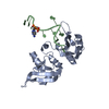 1u1kC 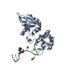 1u1lC 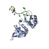 1u1mC 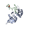 1u1nC 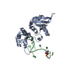 1u1pC  1u1qC 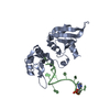 1u1rC 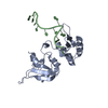 2up1S S: Starting model for refinement C: citing same article ( |
|---|---|
| Similar structure data |


-Links
 PDBj
PDBj


- Assembly
| Deposited unit | 
| ||||||||
|---|---|---|---|---|---|---|---|---|---|
| 1 |
| ||||||||
| Unit cell |
|
-Components
| #1: DNA chain | Mass: 3454.255 Da / Num. of mol.: 1 / Source method: obtained synthetically Details: Oligonucleotide d(TTAGGGTTAG (DI) G) based on human telomeric repeat d(TTAGGG)n |
|---|---|
| #2: Protein | Mass: 22303.082 Da / Num. of mol.: 1 Source method: isolated from a genetically manipulated source Source: (gene. exp.) Homo sapiens (human) / Gene: HNRPA1 / Plasmid: pYS45 / Species (production host): Escherichia coli / Production host:  |
| #3: Water | ChemComp-HOH /  Mass: 18.015 Da / Num. of mol.: 87 / Source method: isolated from a natural source / Formula: H2O Mass: 18.015 Da / Num. of mol.: 87 / Source method: isolated from a natural source / Formula: H2O |
-Experimental details
-Experiment
| Experiment | Method: X-RAY DIFFRACTION / Number of used crystals: 1 |
|---|
- Sample preparation
| Crystal | Density Matthews: 2.248 Å3/Da / Density % sol: 44.26 % | ||||||||||||||||||||||||||||||||||||||||||||||||||||||||||||
|---|---|---|---|---|---|---|---|---|---|---|---|---|---|---|---|---|---|---|---|---|---|---|---|---|---|---|---|---|---|---|---|---|---|---|---|---|---|---|---|---|---|---|---|---|---|---|---|---|---|---|---|---|---|---|---|---|---|---|---|---|---|
| Crystal grow | Temperature: 283 K / Method: vapor diffusion, hanging drop / pH: 8.1 Details: ammonium phosphate, glycerol, Tris, sodium chloride, MES, EDTA, beta-mercaptoethanol, pH 8.1, VAPOR DIFFUSION, HANGING DROP, temperature 283.0K | ||||||||||||||||||||||||||||||||||||||||||||||||||||||||||||
| Components of the solutions |
|
-Data collection
| Diffraction | Mean temperature: 103 K |
|---|---|
| Diffraction source | Source: ROTATING ANODE / Type: RIGAKU RUH3R / Wavelength: 1.5418 Å |
| Detector | Type: RIGAKU RAXIS IV++ / Detector: IMAGE PLATE / Date: Jun 4, 2002 / Details: osmic mirrors |
| Radiation | Protocol: SINGLE WAVELENGTH / Monochromatic (M) / Laue (L): M / Scattering type: x-ray |
| Radiation wavelength | Wavelength: 1.5418 Å / Relative weight: 1 |
| Reflection | Resolution: 2→20 Å / Num. all: 16052 / Num. obs: 15339 / % possible obs: 95.6 % / Observed criterion σ(F): 0 / Observed criterion σ(I): 0 / Redundancy: 6.6 % / Biso Wilson estimate: 28.9 Å2 / Rmerge(I) obs: 0.077 / Rsym value: 0.077 / Net I/σ(I): 12.3 |
| Reflection shell | Resolution: 2→2.07 Å / Redundancy: 2.6 % / Rmerge(I) obs: 0.395 / Mean I/σ(I) obs: 5.1 / Num. unique all: 107 / % possible all: 93.2 |
- Processing
| Software |
| ||||||||||||||||||||||||||||||||||||
|---|---|---|---|---|---|---|---|---|---|---|---|---|---|---|---|---|---|---|---|---|---|---|---|---|---|---|---|---|---|---|---|---|---|---|---|---|---|
| Refinement | Method to determine structure: MOLECULAR REPLACEMENT Starting model: PDB ENTRY 2UP1 Resolution: 2→19.87 Å / Rfactor Rfree error: 0.01 / Data cutoff high absF: 513258.2 / Data cutoff low absF: 0 / Isotropic thermal model: RESTRAINED / Cross valid method: THROUGHOUT / σ(F): 0 / σ(I): 0 / Stereochemistry target values: Engh & Huber
| ||||||||||||||||||||||||||||||||||||
| Solvent computation | Solvent model: FLAT MODEL / Bsol: 39.7392 Å2 / ksol: 0.372048 e/Å3 | ||||||||||||||||||||||||||||||||||||
| Displacement parameters | Biso mean: 33.8 Å2
| ||||||||||||||||||||||||||||||||||||
| Refine analyze |
| ||||||||||||||||||||||||||||||||||||
| Refinement step | Cycle: LAST / Resolution: 2→19.87 Å
| ||||||||||||||||||||||||||||||||||||
| Refine LS restraints |
| ||||||||||||||||||||||||||||||||||||
| LS refinement shell | Resolution: 2→2.07 Å / Rfactor Rfree error: 0.044 / Total num. of bins used: 10
| ||||||||||||||||||||||||||||||||||||
| Xplor file |
|
