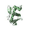
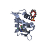
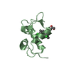
positive regulation of mitotic cell cycle / RING-type E3 ubiquitin transferase / p53 binding / ubiquitin protein ligase activity / regulation of gene expression / ubiquitin-dependent protein catabolic process / protein ubiquitination / apoptotic process / negative regulation of apoptotic process / nucleolus ...positive regulation of mitotic cell cycle / RING-type E3 ubiquitin transferase / p53 binding / ubiquitin protein ligase activity / regulation of gene expression / ubiquitin-dependent protein catabolic process / protein ubiquitination / apoptotic process / negative regulation of apoptotic process / nucleolus / zinc ion binding / nucleoplasm / identical protein binding / nucleus / cytoplasm Similarity search - Function
E3 ubiquitin-protein ligase Mdm2 / MDM2, modified RING finger, HC subclass / MDM2 / SWIB/MDM2 domain / p53 negative regulator Mdm2/Mdm4 / SWIB/MDM2 domain / SWIB/MDM2 domain / SWIB/MDM2 domain profile. / SWIB/MDM2 domain superfamily / Zn-finger in Ran binding protein and others ...E3 ubiquitin-protein ligase Mdm2 / MDM2, modified RING finger, HC subclass / MDM2 / SWIB/MDM2 domain / p53 negative regulator Mdm2/Mdm4 / SWIB/MDM2 domain / SWIB/MDM2 domain / SWIB/MDM2 domain profile. / SWIB/MDM2 domain superfamily / Zn-finger in Ran binding protein and others / Zinc finger, C3HC4 type (RING finger) / Zinc finger RanBP2 type profile. / Zinc finger, RanBP2-type superfamily / Zinc finger RanBP2-type signature. / Zinc finger, RanBP2-type / Zinc finger RING-type profile. / Zinc finger, RING-type / Zinc finger, RING/FYVE/PHD-type / Orthogonal Bundle / Mainly Alpha Similarity search - Domain/homology
In the structure databanks used in Yorodumi, some data are registered as the other names, "COVID-19 virus" and "2019-nCoV". Here are the details of the virus and the list of structure data.
Jan 31, 2019. EMDB accession codes are about to change! (news from PDBe EMDB page)
EMDB accession codes are about to change! (news from PDBe EMDB page)
The allocation of 4 digits for EMDB accession codes will soon come to an end. Whilst these codes will remain in use, new EMDB accession codes will include an additional digit and will expand incrementally as the available range of codes is exhausted. The current 4-digit format prefixed with “EMD-” (i.e. EMD-XXXX) will advance to a 5-digit format (i.e. EMD-XXXXX), and so on. It is currently estimated that the 4-digit codes will be depleted around Spring 2019, at which point the 5-digit format will come into force.
The EM Navigator/Yorodumi systems omit the EMD- prefix.
Related info.:Q: What is EMD? / ID/Accession-code notation in Yorodumi/EM Navigator
Yorodumi is a browser for structure data from EMDB, PDB, SASBDB, etc.
This page is also the successor to EM Navigator detail page, and also detail information page/front-end page for Omokage search.
The word "yorodu" (or yorozu) is an old Japanese word meaning "ten thousand". "mi" (miru) is to see.
Related info.:EMDB / PDB / SASBDB / Comparison of 3 databanks / Yorodumi Search / Aug 31, 2016. New EM Navigator & Yorodumi / Yorodumi Papers / Jmol/JSmol / Function and homology information / Changes in new EM Navigator and Yorodumi
 Movie
Movie Controller

 Yorodumi
 Open data
 Basic information
 Components
 Keywords
 Function and homology information
 X-RAY DIFFRACTION /
  Authors
 Citation
 Structure visualization
 Downloads & links
 Other downloads
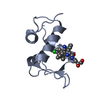


 PDBj
PDBj


 Assembly
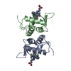


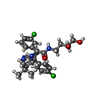
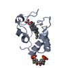
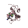
 Mass: 518.475 Da / Num. of mol.: 2 / Source method: obtained synthetically / Formula: C27H33Cl2N3O3
 Mass: 18.015 Da / Num. of mol.: 128 / Source method: isolated from a natural source / Formula: H2O
 Sample preparation
 / Beamline: 31-ID / Type: RIGAKU MICROMAX-007 / Wavelength: 1.5418 Å
 Processing