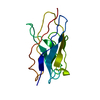
 Movie
Movie Controller


+ Open data

- Basic information
| Entry | Database: PDB / ID: 1t2j | ||||||
|---|---|---|---|---|---|---|---|
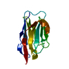
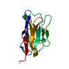
| Title | Crystal structure of a Human VH domain | ||||||
 Components Components | M12-Variable Heavy domain | ||||||
 Keywords Keywords | IMMUNE SYSTEM / beta-sandwich / Immunoglobulin fold | ||||||
| Function / homology |  Function and homology information Function and homology informationimmunoglobulin complex / adaptive immune response / extracellular region / plasma membrane Similarity search - Function | ||||||
| Biological species |  Homo sapiens (human) Homo sapiens (human) | ||||||
| Method |  X-RAY DIFFRACTION / SYNCHROTRON / MOLECULAR REPLACEMENT / Resolution: 1.5 Å X-RAY DIFFRACTION / SYNCHROTRON / MOLECULAR REPLACEMENT / Resolution: 1.5 Å | ||||||
 Authors Authors | Gaur, R.K. / Fischer, R. / Hoffmann, K.M.V. | ||||||
 Citation Citation | Journal: To be Published Title: Crystal structure of a human VH domain at 1.5 angstrom Authors: Gaur, R.K. / Fischer, R. / Hoffmann, K.M.V. | ||||||
| History |
| ||||||
| Remark 999 | SEQUENCE THE AUTHORS STATE THIS SEQUENCE HAS NOT BEEN DEPOSITED TO A SEQUENCE DATABASE. |
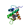
- Structure visualization
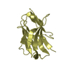
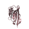
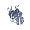
| Structure viewer | Molecule: MolmilJmol/JSmol |
|---|
- Downloads & links
-Download
| PDBx/mmCIF format | 1t2j.cif.gz | 38.7 KB | Display | PDBx/mmCIF format |
|---|---|---|---|---|
| PDB format | pdb1t2j.ent.gz | 26 KB | Display | PDB format |
| PDBx/mmJSON format | 1t2j.json.gz | Tree view | PDBx/mmJSON format | |
| Others |  Other downloads Other downloads |
-Validation report
| Arichive directory | https://data.pdbj.org/pub/pdb/validation_reports/t2/1t2jftp://data.pdbj.org/pub/pdb/validation_reports/t2/1t2j | HTTPS FTP |
|---|
-Related structure data
| Related structure data |  1houS S: Starting model for refinement |
|---|---|
| Similar structure data |

-Links
 PDBj
PDBj

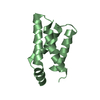
- Assembly
| Deposited unit | 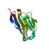
| |||||||||
|---|---|---|---|---|---|---|---|---|---|---|
| 1 |
| |||||||||
| Unit cell |
| |||||||||
| Components on special symmetry positions |
|
-Components
| #1: Antibody | Mass: 12369.602 Da / Num. of mol.: 1 Source method: isolated from a genetically manipulated source Source: (gene. exp.) Homo sapiens (human) / Plasmid: pSyn / Production host:  |
|---|---|
| #2: Chemical | ChemComp-PEG /   Mass: 106.120 Da / Num. of mol.: 1 / Source method: obtained synthetically / Formula: C4H10O3 Mass: 106.120 Da / Num. of mol.: 1 / Source method: obtained synthetically / Formula: C4H10O3 |
| #3: Water | ChemComp-HOH /  Mass: 18.015 Da / Num. of mol.: 139 / Source method: isolated from a natural source / Formula: H2O Mass: 18.015 Da / Num. of mol.: 139 / Source method: isolated from a natural source / Formula: H2O |
| Has protein modification | Y |
-Experimental details
-Experiment
| Experiment | Method: X-RAY DIFFRACTION / Number of used crystals: 1 |
|---|
- Sample preparation
| Crystal | Density Matthews: 1.92 Å3/Da / Density % sol: 35.43 % |
|---|---|
| Crystal grow | Temperature: 277 K / Method: vapor diffusion, hanging drop / pH: 6.5 Details: Crystals grew from 3 microlitre hangingdrop formed by mixing 1.5 microlitre of protein:subtilisin mixture (250:1 M ratio) at protein conc. 13.5 mg/ml with 1.5 microlitre of reservoir ...Details: Crystals grew from 3 microlitre hangingdrop formed by mixing 1.5 microlitre of protein:subtilisin mixture (250:1 M ratio) at protein conc. 13.5 mg/ml with 1.5 microlitre of reservoir solution 100mM MES, pH6.5, 10mM ZnSO4, 25% v/v PEG550MME, 4mM DTT and 0.04% v/v NaN3, VAPOR DIFFUSION, HANGING DROP, temperature 277K |
-Data collection
| Diffraction | Mean temperature: 100 K |
|---|---|
| Diffraction source | Source: SYNCHROTRON / Site: EMBL/DESY, HAMBURG  / Beamline: X31 / Wavelength: 0.802 Å / Beamline: X31 / Wavelength: 0.802 Å |
| Detector | Type: MARRESEARCH / Detector: CCD / Date: Jul 5, 2003 / Details: Rh coated mirror |
| Radiation | Monochromator: Triangular Si (111) / Protocol: SINGLE WAVELENGTH / Monochromatic (M) / Laue (L): M / Scattering type: x-ray |
| Radiation wavelength | Wavelength: 0.802 Å / Relative weight: 1 |
| Reflection | Resolution: 1.5→34.922 Å / Num. obs: 15017 / % possible obs: 90.7 % / Redundancy: 4.29 % / Biso Wilson estimate: 16.67 Å2 / Rmerge(I) obs: 0.047 / Net I/σ(I): 27.69 |
| Reflection shell | Resolution: 1.5→1.53 Å / Redundancy: 4.1 % / Rmerge(I) obs: 0.214 / Mean I/σ(I) obs: 7.26 / Num. unique all: 696 / % possible all: 92.9 |
- Processing
| Software |
| |||||||||||||||||||||||||||||||||||||||||||||||||||||||||||||||||||||||||||
|---|---|---|---|---|---|---|---|---|---|---|---|---|---|---|---|---|---|---|---|---|---|---|---|---|---|---|---|---|---|---|---|---|---|---|---|---|---|---|---|---|---|---|---|---|---|---|---|---|---|---|---|---|---|---|---|---|---|---|---|---|---|---|---|---|---|---|---|---|---|---|---|---|---|---|---|---|
| Refinement | Method to determine structure: MOLECULAR REPLACEMENT Starting model: PDB ENTRY 1HOU Resolution: 1.5→20 Å / Cor.coef. Fo:Fc: 0.973 / Cor.coef. Fo:Fc free: 0.947 / SU B: 1.218 / SU ML: 0.046 / Isotropic thermal model: isotropic / Cross valid method: THROUGHOUT / ESU R: 0.076 / ESU R Free: 0.08 / Stereochemistry target values: MAXIMUM LIKELIHOOD / Details: used weighted full matrix least square procedure
| |||||||||||||||||||||||||||||||||||||||||||||||||||||||||||||||||||||||||||
| Solvent computation | Ion probe radii: 0.8 Å / Shrinkage radii: 0.8 Å / VDW probe radii: 1.4 Å / Solvent model: BABINET MODEL WITH MASK | |||||||||||||||||||||||||||||||||||||||||||||||||||||||||||||||||||||||||||
| Displacement parameters | Biso mean: 21.603 Å2
| |||||||||||||||||||||||||||||||||||||||||||||||||||||||||||||||||||||||||||
| Refine analyze | Luzzati coordinate error obs: 0.153 Å | |||||||||||||||||||||||||||||||||||||||||||||||||||||||||||||||||||||||||||
| Refinement step | Cycle: LAST / Resolution: 1.5→20 Å
| |||||||||||||||||||||||||||||||||||||||||||||||||||||||||||||||||||||||||||
| Refine LS restraints |
| |||||||||||||||||||||||||||||||||||||||||||||||||||||||||||||||||||||||||||
| LS refinement shell | Resolution: 1.5→1.539 Å / Total num. of bins used: 20
|
