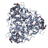
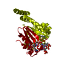
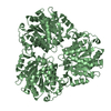
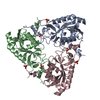
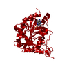
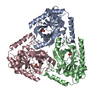
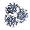
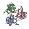
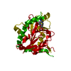
- PDB-4jqs: Crystal structure of a Putative thua-like protein (BACUNI_01602) ... -
+
Open data
ID or keywords:
Loading...
-
Basic information
Entry
Database: PDB / ID: 4jqs
Title
Crystal structure of a Putative thua-like protein (BACUNI_01602) from Bacteroides uniformis ATCC 8492 at 2.30 A resolution
Components
hypothetical protein
Keywords
Structural Genomics / Unknown Function / Trehalose utilization / PF06283 family protein / Joint Center for Structural Genomics / JCSG / Protein Structure Initiative / PSI-BIOLOGY
Function / homology
ThuA-like domain / Trehalose utilisation / Class I glutamine amidotransferase (GATase) domain / Class I glutamine amidotransferase-like / Rossmann fold / 3-Layer(aba) Sandwich / metal ion binding / Alpha Beta / ThuA-like domain-containing protein
Function and homology information
Biological species
Bacteroides uniformis (bacteria)
Method
X-RAY DIFFRACTION / SYNCHROTRON / MAD / Resolution: 2.3 Å
Mass: 18.015 Da / Num. of mol.: 258 / Source method: isolated from a natural source / Formula: H2O
Has protein modification
Y
Sequence details
THE CONSTRUCT (RESIDUES 19-267) WAS EXPRESSED WITH A PURIFICATION TAG MGSDKIHHHHHHENLYFQG. THE TAG ...THE CONSTRUCT (RESIDUES 19-267) WAS EXPRESSED WITH A PURIFICATION TAG MGSDKIHHHHHHENLYFQG. THE TAG WAS REMOVED WITH TEV PROTEASE LEAVING ONLY A GLYCINE (0) FOLLOWED BY THE TARGET SEQUENCE.
-
Experimental details
-
Experiment
Experiment
Method: X-RAY DIFFRACTION / Number of used crystals: 1
-
Sample preparation
Crystal
Density Matthews: 2.28 Å3/Da / Density % sol: 46 %
Crystal grow
Temperature: 293 K / Method: vapor diffusion, sitting drop / pH: 9.5 Details: 0.1M CHES pH 9.5, 40% polyethylene glycol 600, NANODROP, VAPOR DIFFUSION, SITTING DROP, temperature 293K
Type: DECTRIS PILATUS 6M / Detector: PIXEL / Date: Jan 23, 2013 Details: Flat mirror (vertical focusing); single crystal Si(111) bent monochromator (horizontal focusing)
Radiation
Monochromator: single crystal Si(111) bent / Protocol: MAD / Monochromatic (M) / Laue (L): M / Scattering type: x-ray
Radiation wavelength
ID
Wavelength (Å)
Relative weight
1
0.91837
1
2
0.97935
1
3
0.97879
1
Reflection
Resolution: 2.3→28.834 Å / Num. obs: 35202 / % possible obs: 97 % / Observed criterion σ(I): -3 / Biso Wilson estimate: 50.79 Å2 / Rmerge(I) obs: 0.064 / Net I/σ(I): 11.63
Reflection shell
Resolution (Å)
Highest resolution (Å)
Rmerge(I) obs
Mean I/σ(I) obs
Diffraction-ID
% possible all
2.3-2.38
0.629
1.4
1
91
2.38-2.48
0.489
1.9
1
98.7
2.48-2.59
0.365
2.6
1
99.1
2.59-2.73
0.259
3.6
1
98.9
2.73-2.9
0.183
5
1
97.8
2.9-3.12
0.133
6.9
1
93.5
3.12-3.43
0.071
12.3
1
99.3
3.43-3.93
0.042
19.7
1
98.7
3.93-4.93
0.027
28.2
1
95.5
4.93
0.022
34.2
1
97.3
-
Phasing
Phasing
Method: MAD
-
Processing
Software
Name
Version
Classification
NB
MolProbity
3beta29
modelbuilding
PDB_EXTRACT
3.1
dataextraction
SOLVE
phasing
XSCALE
July4, 2012
datascaling
BUSTER-TNT
2.10.0
refinement
XDS
datareduction
BUSTER
2.10.0
refinement
Refinement
Method to determine structure: MAD / Resolution: 2.3→28.09 Å / Cor.coef. Fo:Fc: 0.959 / Cor.coef. Fo:Fc free: 0.9285 / Occupancy max: 1 / Occupancy min: 0.5 / Cross valid method: THROUGHOUT / σ(F): 0 Details: 1. A MET-INHIBITION PROTOCOL WAS USED FOR SELENOMETHIONINE INCORPORATION DURING PROTEIN EXPRESSION. THE OCCUPANCY OF THE SE ATOMS IN THE MSE RESIDUES WAS REDUCED TO 0.75 FOR THE REDUCED ...Details: 1. A MET-INHIBITION PROTOCOL WAS USED FOR SELENOMETHIONINE INCORPORATION DURING PROTEIN EXPRESSION. THE OCCUPANCY OF THE SE ATOMS IN THE MSE RESIDUES WAS REDUCED TO 0.75 FOR THE REDUCED SCATTERING POWER DUE TO PARTIAL S-MET INCORPORATION. 2. ATOM RECORD CONTAINS SUM OF TLS AND RESIDUAL B FACTORS. ANISOU RECORD CONTAINS SUM OF TLS AND RESIDUAL U FACTORS. 3. THE REFINEMENT WAS RESTRAINED AGAINST THE MAD PHASES. 4. NCS RESTRAINTS WERE APPLIED USING BUSTER'S LSSR RESTRAINT REPRESENTATION (-AUTONCS). 5. POLYETHYLENE GLYCOL FRAGMENTS (PG4,P6G) FROM THE CRYSTALLIZATION SOLUTION WERE MODELED INTO THE STRUCTURE.
Rfactor
Num. reflection
% reflection
Selection details
Rfree
0.2061
1764
5.01 %
RANDOM
Rwork
0.1623
-
-
-
obs
0.1645
35175
98.91 %
-
Displacement parameters
Biso mean: 43.84 Å2
Baniso -1
Baniso -2
Baniso -3
1-
2.7367 Å2
0 Å2
0 Å2
2-
-
-1.4678 Å2
0 Å2
3-
-
-
-1.2689 Å2
Refine analyze
Luzzati coordinate error obs: 0.266 Å
Refinement step
Cycle: LAST / Resolution: 2.3→28.09 Å
Protein
Nucleic acid
Ligand
Solvent
Total
Num. atoms
5996
0
45
258
6299
Refine LS restraints
Refine-ID
Type
Dev ideal
Number
Restraint function
Weight
X-RAY DIFFRACTION
t_bond_d
0.01
6258
HARMONIC
2
X-RAY DIFFRACTION
t_angle_deg
0.98
8520
HARMONIC
2
X-RAY DIFFRACTION
t_dihedral_angle_d
2728
SINUSOIDAL
2
X-RAY DIFFRACTION
t_incorr_chiral_ct
X-RAY DIFFRACTION
t_pseud_angle
X-RAY DIFFRACTION
t_trig_c_planes
170
HARMONIC
2
X-RAY DIFFRACTION
t_gen_planes
887
HARMONIC
5
X-RAY DIFFRACTION
t_it
6258
HARMONIC
20
X-RAY DIFFRACTION
t_nbd
X-RAY DIFFRACTION
t_omega_torsion
3.6
X-RAY DIFFRACTION
t_other_torsion
17.59
X-RAY DIFFRACTION
t_improper_torsion
X-RAY DIFFRACTION
t_chiral_improper_torsion
745
SEMIHARMONIC
5
X-RAY DIFFRACTION
t_sum_occupancies
X-RAY DIFFRACTION
t_utility_distance
X-RAY DIFFRACTION
t_utility_angle
X-RAY DIFFRACTION
t_utility_torsion
X-RAY DIFFRACTION
t_ideal_dist_contact
7337
SEMIHARMONIC
4
LS refinement shell
Resolution: 2.3→2.37 Å / Total num. of bins used: 18
Rfactor
Num. reflection
% reflection
Rfree
0.248
127
4.61 %
Rwork
0.2088
2626
-
all
0.2106
2753
-
obs
-
-
98.91 %
Refinement TLS params.
Method: refined / Refine-ID: X-RAY DIFFRACTION
ID
L11 (°2)
L12 (°2)
L13 (°2)
L22 (°2)
L23 (°2)
L33 (°2)
S11 (Å °)
S12 (Å °)
S13 (Å °)
S21 (Å °)
S22 (Å °)
S23 (Å °)
S31 (Å °)
S32 (Å °)
S33 (Å °)
T11 (Å2)
T12 (Å2)
T13 (Å2)
T22 (Å2)
T23 (Å2)
T33 (Å2)
Origin x (Å)
Origin y (Å)
Origin z (Å)
1
1.4592
0.1105
-0.1676
1.0804
-0.5117
1.2651
-0.0108
0.1475
-0.2312
-0.1685
0.0914
-0.0678
0.3017
0.0477
-0.0806
0.0132
-0.01
-0.0292
-0.1054
-0.0647
-0.084
43.3835
69.9774
90.2465
2
1.5621
-0.1118
0.597
1.0118
-0.1675
0.9502
-0.0375
0.2327
0.2809
-0.0283
0.0401
-0.1711
-0.0992
0.193
-0.0025
-0.0788
-0.0603
0.0268
-0.0478
0.0584
-0.0352
54.1586
105.222
89.3735
3
1.1939
0.0297
-0.2133
0.8366
0.4329
1.1787
-0.002
0.0891
0.1747
-0.0488
0.0587
0.2227
-0.0577
-0.146
-0.0567
-0.0864
-0.0068
-0.0387
-0.0505
0.0827
-0.0146
18.2317
96.9931
92.4199
Refinement TLS group
ID
Refine-ID
Refine TLS-ID
Selection details
Auth asym-ID
Auth seq-ID
1
X-RAY DIFFRACTION
1
{ A|23 - 267 }
A
23 - 267
2
X-RAY DIFFRACTION
2
{ B|23 - 267 }
B
23 - 267
3
X-RAY DIFFRACTION
3
{ C|23 - 267 }
C
23 - 267
+
About Yorodumi
-
News
-
Feb 9, 2022. New format data for meta-information of EMDB entries
New format data for meta-information of EMDB entries
Version 3 of the EMDB header file is now the official format.
The previous official version 1.9 will be removed from the archive.
In the structure databanks used in Yorodumi, some data are registered as the other names, "COVID-19 virus" and "2019-nCoV". Here are the details of the virus and the list of structure data.
Jan 31, 2019. EMDB accession codes are about to change! (news from PDBe EMDB page)
EMDB accession codes are about to change! (news from PDBe EMDB page)
The allocation of 4 digits for EMDB accession codes will soon come to an end. Whilst these codes will remain in use, new EMDB accession codes will include an additional digit and will expand incrementally as the available range of codes is exhausted. The current 4-digit format prefixed with “EMD-” (i.e. EMD-XXXX) will advance to a 5-digit format (i.e. EMD-XXXXX), and so on. It is currently estimated that the 4-digit codes will be depleted around Spring 2019, at which point the 5-digit format will come into force.
The EM Navigator/Yorodumi systems omit the EMD- prefix.
Related info.:Q: What is EMD? / ID/Accession-code notation in Yorodumi/EM Navigator
Yorodumi is a browser for structure data from EMDB, PDB, SASBDB, etc.
This page is also the successor to EM Navigator detail page, and also detail information page/front-end page for Omokage search.
The word "yorodu" (or yorozu) is an old Japanese word meaning "ten thousand". "mi" (miru) is to see.
Related info.:EMDB / PDB / SASBDB / Comparison of 3 databanks / Yorodumi Search / Aug 31, 2016. New EM Navigator & Yorodumi / Yorodumi Papers / Jmol/JSmol / Function and homology information / Changes in new EM Navigator and Yorodumi
 Movie
Movie Controller

 Yorodumi
 Open data
 Basic information
 Components
 Keywords
 Function and homology information
 Bacteroides uniformis (bacteria)
 X-RAY DIFFRACTION /
  Authors
 Citation
 Structure visualization
 Downloads & links
 Other downloads
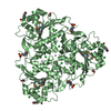
 PDBj
PDBj

 Assembly
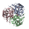

 Mass: 194.226 Da / Num. of mol.: 2 / Source method: obtained synthetically / Formula: C8H18O5 / Comment: precipitant*YM
 Mass: 282.331 Da / Num. of mol.: 1 / Source method: obtained synthetically / Formula: C12H26O7 / Comment: precipitant*YM
 Mass: 18.015 Da / Num. of mol.: 258 / Source method: isolated from a natural source / Formula: H2O
 Sample preparation
 / Beamline: BL11-1 / Wavelength: 0.91837,0.97935,0.97879
 Processing