 Movie
Movie Controller

[English] 日本語
 Yorodumi
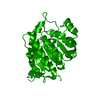
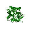
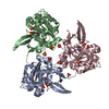
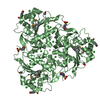
- PDB-4mxr: Crystal structure of Trypanosoma cruzi formiminoglutamase with Mn2+2 -
+ Open data

- Basic information
| Entry | Database: PDB / ID: 4mxr | ||||||
|---|---|---|---|---|---|---|---|
| Title | Crystal structure of Trypanosoma cruzi formiminoglutamase with Mn2+2 | ||||||
 Components Components | Formiminoglutamase | ||||||
 Keywords Keywords | HYDROLASE / Arginase/deacetylase (a/b) fold | ||||||
| Function / homology |  Function and homology information Function and homology information | ||||||
| Biological species |  | ||||||
| Method |  X-RAY DIFFRACTION / MOLECULAR REPLACEMENT / Resolution: 1.849 Å X-RAY DIFFRACTION / MOLECULAR REPLACEMENT / Resolution: 1.849 Å | ||||||
 Authors Authors | Hai, Y. / Dugery, R.-J. / Healy, D. / Christianson, D.W. | ||||||
 Citation Citation | Journal: Biochemistry / Year: 2013 Title: Formiminoglutamase from trypanosoma cruzi is an arginase-like manganese metalloenzyme. Authors: Hai, Y. / Dugery, R.J. / Healy, D. / Christianson, D.W. | ||||||
| History |
|
- Structure visualization
| Structure viewer | Molecule: MolmilJmol/JSmol |
|---|
- Downloads & links
-Download
| PDBx/mmCIF format | 4mxr.cif.gz | 132.4 KB | Display | PDBx/mmCIF format |
|---|---|---|---|---|
| PDB format | pdb4mxr.ent.gz | 101.8 KB | Display | PDB format |
| PDBx/mmJSON format | 4mxr.json.gz | Tree view | PDBx/mmJSON format | |
| Others |  Other downloads Other downloads |
-Validation report
| Arichive directory | https://data.pdbj.org/pub/pdb/validation_reports/mx/4mxrftp://data.pdbj.org/pub/pdb/validation_reports/mx/4mxr | HTTPS FTP |
|---|
-Related structure data
| Related structure data | 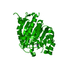 4myfC 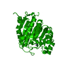 4mykC  4mylC  4mynC 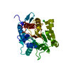 2a0mS S: Starting model for refinement C: citing same article ( |
|---|---|
| Similar structure data |


-Links
 PDBj
PDBj- Assembly
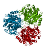
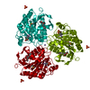
| Deposited unit | 
| |||||||||
|---|---|---|---|---|---|---|---|---|---|---|
| 1 | 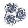
| |||||||||
| 2 | 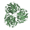
| |||||||||
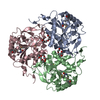
| Unit cell |
| |||||||||
| Components on special symmetry positions |
|
-Components
| #1: Protein | Mass: 34637.227 Da / Num. of mol.: 2 / Mutation: S302P Source method: isolated from a genetically manipulated source Source: (gene. exp.)  #2: Chemical | ChemComp-MN /   Mass: 54.938 Da / Num. of mol.: 4 / Source method: obtained synthetically / Formula: Mn Mass: 54.938 Da / Num. of mol.: 4 / Source method: obtained synthetically / Formula: Mn#3: Chemical | ChemComp-GOL / |   Mass: 92.094 Da / Num. of mol.: 1 / Source method: obtained synthetically / Formula: C3H8O3 Mass: 92.094 Da / Num. of mol.: 1 / Source method: obtained synthetically / Formula: C3H8O3#4: Water | ChemComp-HOH / |  Mass: 18.015 Da / Num. of mol.: 265 / Source method: isolated from a natural source / Formula: H2O Mass: 18.015 Da / Num. of mol.: 265 / Source method: isolated from a natural source / Formula: H2O |
|---|
-Experimental details
-Experiment
| Experiment | Method: X-RAY DIFFRACTION / Number of used crystals: 1 |
|---|
- Sample preparation
| Crystal | Density Matthews: 1.97 Å3/Da / Density % sol: 37.6 % |
|---|---|
| Crystal grow | Temperature: 294 K / Method: vapor diffusion, hanging drop / pH: 4.9 Details: A 4 UL drop of protein solution [10 mg/mL protein, 50 mM bicine (pH 8.5), 100 UM MnCl2, 1 mM TCEP] was mixed with a 4 UL drop of precipitant solution [31% PEG 300, 0.1 M sodium acetate (pH 4. ...Details: A 4 UL drop of protein solution [10 mg/mL protein, 50 mM bicine (pH 8.5), 100 UM MnCl2, 1 mM TCEP] was mixed with a 4 UL drop of precipitant solution [31% PEG 300, 0.1 M sodium acetate (pH 4.9)] on a siliconized cover slide and equilibrated against a 500 UL reservoir of precipitant solution at 294K. The yielded crystal is apo. To obtain the Mn2+2 bound form, this apo-crystal was soaked with 5 mM MnCl2 in 0.1 M sodium malonate (pH 8.0), 27% PEG 3350) for 24 hours, VAPOR DIFFUSION, HANGING DROP |
-Data collection
| Diffraction | Mean temperature: 100 K |
|---|---|
| Diffraction source | Source: ROTATING ANODE / Type: RIGAKU RU200 / Wavelength: 1.5418 Å |
| Detector | Type: RIGAKU RAXIS IV++ / Detector: IMAGE PLATE / Date: Feb 17, 2013 / Details: mirrors |
| Radiation | Monochromator: Yale Mirrors / Protocol: SINGLE WAVELENGTH / Monochromatic (M) / Laue (L): M / Scattering type: x-ray |
| Radiation wavelength | Wavelength: 1.5418 Å / Relative weight: 1 |
| Reflection | Resolution: 1.85→50 Å / Num. obs: 44906 / % possible obs: 99.2 % / Observed criterion σ(F): 0 / Observed criterion σ(I): -3 / Redundancy: 2.5 % / Rmerge(I) obs: 0.059 / Rsym value: 0.059 / Net I/σ(I): 14.162 |
| Reflection shell | Resolution: 1.85→1.92 Å / Redundancy: 2.4 % / Rmerge(I) obs: 0.438 / Mean I/σ(I) obs: 2.013 / Rsym value: 0.438 / % possible all: 98.8 |
- Processing
| Software |
| ||||||||||||||||||||||||||||||||||||||||||||||||||||||||||||||||||||||||||||||||||||||||||||||||||||||||||||||||
|---|---|---|---|---|---|---|---|---|---|---|---|---|---|---|---|---|---|---|---|---|---|---|---|---|---|---|---|---|---|---|---|---|---|---|---|---|---|---|---|---|---|---|---|---|---|---|---|---|---|---|---|---|---|---|---|---|---|---|---|---|---|---|---|---|---|---|---|---|---|---|---|---|---|---|---|---|---|---|---|---|---|---|---|---|---|---|---|---|---|---|---|---|---|---|---|---|---|---|---|---|---|---|---|---|---|---|---|---|---|---|---|---|---|
| Refinement | Method to determine structure: MOLECULAR REPLACEMENT Starting model: PDB ENTRY 2A0M Resolution: 1.849→21.964 Å / SU ML: 0.22 / Cross valid method: THROUGHOUT / σ(F): 0.06 / Phase error: 27 / Stereochemistry target values: ML
| ||||||||||||||||||||||||||||||||||||||||||||||||||||||||||||||||||||||||||||||||||||||||||||||||||||||||||||||||
| Solvent computation | Shrinkage radii: 0.9 Å / VDW probe radii: 1.11 Å / Solvent model: FLAT BULK SOLVENT MODEL / Bsol: 40.001 Å2 / ksol: 0.35 e/Å3 | ||||||||||||||||||||||||||||||||||||||||||||||||||||||||||||||||||||||||||||||||||||||||||||||||||||||||||||||||
| Displacement parameters |
| ||||||||||||||||||||||||||||||||||||||||||||||||||||||||||||||||||||||||||||||||||||||||||||||||||||||||||||||||
| Refinement step | Cycle: LAST / Resolution: 1.849→21.964 Å
| ||||||||||||||||||||||||||||||||||||||||||||||||||||||||||||||||||||||||||||||||||||||||||||||||||||||||||||||||
| Refine LS restraints |
| ||||||||||||||||||||||||||||||||||||||||||||||||||||||||||||||||||||||||||||||||||||||||||||||||||||||||||||||||
| LS refinement shell |
|
