 Movie
Movie Controller

[English] 日本語
 Yorodumi
- PDB-4jqd: Crystal structure of the Restriction-Modification Controller Prot... -
+ Open data

- Basic information
| Entry | Database: PDB / ID: 4jqd | ||||||
|---|---|---|---|---|---|---|---|
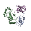
| Title | Crystal structure of the Restriction-Modification Controller Protein C.Csp231I OL operator complex | ||||||
 Components Components |
| ||||||
 Keywords Keywords | TRANSCRIPTION/DNA / helix-turn-helix / C controller protein / restriction-modification systems / transcriptional regulation / TRANSCRIPTION-DNA complex | ||||||
| Function / homology |  Function and homology information Function and homology information | ||||||
| Biological species |  Citrobacter sp. RFL231 (bacteria) Citrobacter sp. RFL231 (bacteria) | ||||||
| Method |  X-RAY DIFFRACTION / SYNCHROTRON / MOLECULAR REPLACEMENT / Resolution: 2.75 Å X-RAY DIFFRACTION / SYNCHROTRON / MOLECULAR REPLACEMENT / Resolution: 2.75 Å | ||||||
 Authors Authors | Shevtsov, M.B. / Streeter, S.D. / Thresh, S.J. / McGeehan, J.E. / Kneale, G.G. | ||||||
 Citation Citation | Journal: Acta Crystallogr.,Sect.D / Year: 2015 Title: Structural analysis of DNA binding by C.Csp231I, a member of a novel class of R-M controller proteins regulating gene expression. Authors: Shevtsov, M.B. / Streeter, S.D. / Thresh, S.J. / Swiderska, A. / McGeehan, J.E. / Kneale, G.G. | ||||||
| History |
|
- Structure visualization
| Structure viewer | Molecule: MolmilJmol/JSmol |
|---|
- Downloads & links
-Download
| PDBx/mmCIF format | 4jqd.cif.gz | 254.6 KB | Display | PDBx/mmCIF format |
|---|---|---|---|---|
| PDB format | pdb4jqd.ent.gz | 203.9 KB | Display | PDB format |
| PDBx/mmJSON format | 4jqd.json.gz | Tree view | PDBx/mmJSON format | |
| Others |  Other downloads Other downloads |
-Validation report
| Arichive directory | https://data.pdbj.org/pub/pdb/validation_reports/jq/4jqdftp://data.pdbj.org/pub/pdb/validation_reports/jq/4jqd | HTTPS FTP |
|---|
-Related structure data
| Related structure data | 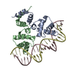 4jcxC 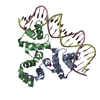 4jcyC  3lfpS S: Starting model for refinement C: citing same article ( |
|---|---|
| Similar structure data |


-Links
 PDBj
PDBj


- Assembly
| Deposited unit | 
| ||||||||
|---|---|---|---|---|---|---|---|---|---|
| 1 | 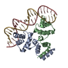
| ||||||||
| 2 | 
| ||||||||
| Unit cell |
| ||||||||
| Details | AUTHORS STATE THAT THE BIOLOGICAL ASSEMBLY IS A PROTEIN DIMER BOUND TO DNA DUPLEX. THE ASYMMETRIC UNIT CONTAINS TWO PROTEIN-DNA COMPLEXES. |
-Components
| #1: Protein | Mass: 11380.236 Da / Num. of mol.: 4 Source method: isolated from a genetically manipulated source Source: (gene. exp.) Citrobacter sp. RFL231 (bacteria) / Plasmid: pET-11a / Production host: #2: DNA chain | Mass: 6472.251 Da / Num. of mol.: 2 / Source method: obtained synthetically #3: DNA chain | Mass: 6409.153 Da / Num. of mol.: 2 / Source method: obtained synthetically #4: Water | ChemComp-HOH / |  Mass: 18.015 Da / Num. of mol.: 51 / Source method: isolated from a natural source / Formula: H2O Mass: 18.015 Da / Num. of mol.: 51 / Source method: isolated from a natural source / Formula: H2O |
|---|
-Experimental details
-Experiment
| Experiment | Method: X-RAY DIFFRACTION / Number of used crystals: 1 |
|---|
- Sample preparation
| Crystal | Density Matthews: 2.85 Å3/Da / Density % sol: 56.89 % |
|---|---|
| Crystal grow | Temperature: 289 K / Method: vapor diffusion, sitting drop Details: Protein was dialysed against the buffer containing 0.1 M NaCl, 50 mM TRIS-HCl pH 8.2, 1 mM DTT, and 1 mM EDTA. Crystallisation conditions: 0.2 M sodium nitrate, 0.1 M Bis-Tris-Propane pH 7. ...Details: Protein was dialysed against the buffer containing 0.1 M NaCl, 50 mM TRIS-HCl pH 8.2, 1 mM DTT, and 1 mM EDTA. Crystallisation conditions: 0.2 M sodium nitrate, 0.1 M Bis-Tris-Propane pH 7.5, 24 % (w/v) PEG3350, protein dimer/DNA molar ratio 1:1, protein concentration 1.46 mg/ml, VAPOR DIFFUSION, SITTING DROP, temperature 289K |
-Data collection
| Diffraction | Mean temperature: 120 K |
|---|---|
| Diffraction source | Source: SYNCHROTRON / Site: ESRF  / Beamline: ID14-4 / Wavelength: 0.9393 Å / Beamline: ID14-4 / Wavelength: 0.9393 Å |
| Detector | Type: ADSC QUANTUM 315r / Detector: CCD / Date: Nov 23, 2010 |
| Radiation | Protocol: SINGLE WAVELENGTH / Monochromatic (M) / Laue (L): M / Scattering type: x-ray |
| Radiation wavelength | Wavelength: 0.9393 Å / Relative weight: 1 |
| Reflection | Resolution: 2.75→36 Å / Num. all: 20836 / Num. obs: 20336 / % possible obs: 97.6 % / Observed criterion σ(I): 2 / Redundancy: 3.9 % / Biso Wilson estimate: 43.3 Å2 / Rmerge(I) obs: 0.075 / Net I/σ(I): 9 |
| Reflection shell | Resolution: 2.75→2.94 Å / Redundancy: 3.1 % / Rmerge(I) obs: 0.304 / Mean I/σ(I) obs: 2.9 / Num. unique all: 3378 / % possible all: 89 |
- Processing
| Software |
| |||||||||||||||||||||||||||||||||||||||||||||||||||||||||||||||||||||||||||||||||||||||||||||||||||||||||||||||||||||||||||||||||||||||||||||||||||||||||||||||||||||||||||||||
|---|---|---|---|---|---|---|---|---|---|---|---|---|---|---|---|---|---|---|---|---|---|---|---|---|---|---|---|---|---|---|---|---|---|---|---|---|---|---|---|---|---|---|---|---|---|---|---|---|---|---|---|---|---|---|---|---|---|---|---|---|---|---|---|---|---|---|---|---|---|---|---|---|---|---|---|---|---|---|---|---|---|---|---|---|---|---|---|---|---|---|---|---|---|---|---|---|---|---|---|---|---|---|---|---|---|---|---|---|---|---|---|---|---|---|---|---|---|---|---|---|---|---|---|---|---|---|---|---|---|---|---|---|---|---|---|---|---|---|---|---|---|---|---|---|---|---|---|---|---|---|---|---|---|---|---|---|---|---|---|---|---|---|---|---|---|---|---|---|---|---|---|---|---|---|---|---|
| Refinement | Method to determine structure: MOLECULAR REPLACEMENT Starting model: PDB entry 3LFP Resolution: 2.75→35.06 Å / Cor.coef. Fo:Fc: 0.957 / Cor.coef. Fo:Fc free: 0.936 / SU B: 29.628 / SU ML: 0.267 / Isotropic thermal model: isotropic / Cross valid method: THROUGHOUT / ESU R Free: 0.333 / Stereochemistry target values: MAXIMUM LIKELIHOOD
| |||||||||||||||||||||||||||||||||||||||||||||||||||||||||||||||||||||||||||||||||||||||||||||||||||||||||||||||||||||||||||||||||||||||||||||||||||||||||||||||||||||||||||||||
| Solvent computation | Ion probe radii: 0.8 Å / Shrinkage radii: 0.8 Å / VDW probe radii: 1.2 Å / Solvent model: MASK | |||||||||||||||||||||||||||||||||||||||||||||||||||||||||||||||||||||||||||||||||||||||||||||||||||||||||||||||||||||||||||||||||||||||||||||||||||||||||||||||||||||||||||||||
| Displacement parameters | Biso mean: 50.112 Å2
| |||||||||||||||||||||||||||||||||||||||||||||||||||||||||||||||||||||||||||||||||||||||||||||||||||||||||||||||||||||||||||||||||||||||||||||||||||||||||||||||||||||||||||||||
| Refinement step | Cycle: LAST / Resolution: 2.75→35.06 Å
| |||||||||||||||||||||||||||||||||||||||||||||||||||||||||||||||||||||||||||||||||||||||||||||||||||||||||||||||||||||||||||||||||||||||||||||||||||||||||||||||||||||||||||||||
| Refine LS restraints |
| |||||||||||||||||||||||||||||||||||||||||||||||||||||||||||||||||||||||||||||||||||||||||||||||||||||||||||||||||||||||||||||||||||||||||||||||||||||||||||||||||||||||||||||||
| LS refinement shell | Resolution: 2.75→2.821 Å / Total num. of bins used: 20
| |||||||||||||||||||||||||||||||||||||||||||||||||||||||||||||||||||||||||||||||||||||||||||||||||||||||||||||||||||||||||||||||||||||||||||||||||||||||||||||||||||||||||||||||
| Refinement TLS params. | Method: refined / Refine-ID: X-RAY DIFFRACTION
| |||||||||||||||||||||||||||||||||||||||||||||||||||||||||||||||||||||||||||||||||||||||||||||||||||||||||||||||||||||||||||||||||||||||||||||||||||||||||||||||||||||||||||||||
| Refinement TLS group |
|
