 Movie
Movie Controller

[English] 日本語
 Yorodumi
- PDB-3lfp: Crystal Structure of the Restriction-Modification Controller Prot... -
+ Open data

- Basic information
| Entry | Database: PDB / ID: 3lfp | ||||||
|---|---|---|---|---|---|---|---|
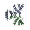
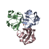
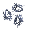
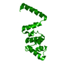
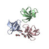
| Title | Crystal Structure of the Restriction-Modification Controller Protein C.Csp231I | ||||||
 Components Components | Csp231I C protein | ||||||
 Keywords Keywords | TRANSCRIPTION / transcriptional regulator / DNA binding protein / helix-turn-helix / restriction-modification | ||||||
| Function / homology |  Function and homology information Function and homology information | ||||||
| Biological species |  Citrobacter sp. RFL231 (bacteria) Citrobacter sp. RFL231 (bacteria) | ||||||
| Method |  X-RAY DIFFRACTION / SYNCHROTRON / MOLECULAR REPLACEMENT / molecular replacement / Resolution: 2 Å X-RAY DIFFRACTION / SYNCHROTRON / MOLECULAR REPLACEMENT / molecular replacement / Resolution: 2 Å | ||||||
 Authors Authors | McGeehan, J.E. / Streeter, S.D. / Thresh, S.J. / Kneale, G.G. | ||||||
 Citation Citation | Journal: J.Mol.Biol. / Year: 2011 Title: Structural Analysis of a Novel Class of R-M Controller Proteins: C.Csp231I from Citrobacter sp. RFL231. Authors: McGeehan, J.E. / Streeter, S.D. / Thresh, S.J. / Taylor, J.E. / Shevtsov, M.B. / Kneale, G.G. | ||||||
| History |
|
- Structure visualization
| Structure viewer | Molecule: MolmilJmol/JSmol |
|---|
- Downloads & links
-Download
| PDBx/mmCIF format | 3lfp.cif.gz | 32.7 KB | Display | PDBx/mmCIF format |
|---|---|---|---|---|
| PDB format | pdb3lfp.ent.gz | 23.8 KB | Display | PDB format |
| PDBx/mmJSON format | 3lfp.json.gz | Tree view | PDBx/mmJSON format | |
| Others |  Other downloads Other downloads |
-Validation report
| Arichive directory | https://data.pdbj.org/pub/pdb/validation_reports/lf/3lfpftp://data.pdbj.org/pub/pdb/validation_reports/lf/3lfp | HTTPS FTP |
|---|
-Related structure data
| Related structure data |  3lisC  1y7yS S: Starting model for refinement C: citing same article ( |
|---|---|
| Similar structure data |


-Links
 PDBj
PDBj


- Assembly
| Deposited unit | 
| ||||||||
|---|---|---|---|---|---|---|---|---|---|
| 1 | 
| ||||||||
| Unit cell |
| ||||||||
| Components on special symmetry positions |
|
-Components
| #1: Protein | Mass: 11380.236 Da / Num. of mol.: 1 Source method: isolated from a genetically manipulated source Source: (gene. exp.) Citrobacter sp. RFL231 (bacteria) / Gene: csp231IC / Plasmid: pET11a / Production host: |
|---|---|
| #2: Water | ChemComp-HOH /  Mass: 18.015 Da / Num. of mol.: 64 / Source method: isolated from a natural source / Formula: H2O Mass: 18.015 Da / Num. of mol.: 64 / Source method: isolated from a natural source / Formula: H2O |
-Experimental details
-Experiment
| Experiment | Method: X-RAY DIFFRACTION / Number of used crystals: 1 |
|---|
- Sample preparation
| Crystal | Density Matthews: 2.37 Å3/Da / Density % sol: 48.16 % |
|---|---|
| Crystal grow | Temperature: 289 K / Method: vapor diffusion, hanging drop / pH: 7.5 Details: 0.1M Na HEPES, 1.4M Tri-sodium citrate dihydrate, pH 7.5, VAPOR DIFFUSION, HANGING DROP, temperature 289K |
-Data collection
| Diffraction | Mean temperature: 100 K |
|---|---|
| Diffraction source | Source: SYNCHROTRON / Site: Diamond  / Beamline: I02 / Wavelength: 0.9795 Å / Beamline: I02 / Wavelength: 0.9795 Å |
| Detector | Type: ADSC QUANTUM 315 / Detector: CCD / Date: Oct 26, 2009 |
| Radiation | Monochromator: Si (111) double crystal monochromator / Protocol: SINGLE WAVELENGTH / Monochromatic (M) / Laue (L): M / Scattering type: x-ray |
| Radiation wavelength | Wavelength: 0.9795 Å / Relative weight: 1 |
| Reflection | Resolution: 2→50 Å / Num. all: 7979 / Num. obs: 7753 / % possible obs: 97.2 % / Observed criterion σ(I): 0 / Redundancy: 38.7 % / Biso Wilson estimate: 35.293 Å2 / Rmerge(I) obs: 0.072 / Net I/σ(I): 42.78 |
| Reflection shell | Resolution: 2→2.1 Å / Redundancy: 38.8 % / Rmerge(I) obs: 0.408 / Mean I/σ(I) obs: 11.3 / Num. measured obs: 42928 / Num. unique all: 991 / Num. unique obs: 1052 / % possible all: 94.2 |
-Phasing
| Phasing | Method: molecular replacement |
|---|
- Processing
| Software |
| |||||||||||||||||||||||||||||||||||||||||||||||||||||||||||||||||
|---|---|---|---|---|---|---|---|---|---|---|---|---|---|---|---|---|---|---|---|---|---|---|---|---|---|---|---|---|---|---|---|---|---|---|---|---|---|---|---|---|---|---|---|---|---|---|---|---|---|---|---|---|---|---|---|---|---|---|---|---|---|---|---|---|---|---|
| Refinement | Method to determine structure: MOLECULAR REPLACEMENT Starting model: PDB Entry 1Y7Y Resolution: 2→19.24 Å / Cor.coef. Fo:Fc: 0.958 / Cor.coef. Fo:Fc free: 0.933 / WRfactor Rfree: 0.226 / WRfactor Rwork: 0.172 / Occupancy max: 1 / Occupancy min: 0.33 / FOM work R set: 0.871 / SU B: 2.794 / SU ML: 0.083 / SU R Cruickshank DPI: 0.162 / SU Rfree: 0.154 / Cross valid method: THROUGHOUT / σ(F): 0 / ESU R: 0.162 / ESU R Free: 0.154 / Stereochemistry target values: MAXIMUM LIKELIHOOD Details: HYDROGENS HAVE BEEN ADDED IN THE RIDING POSITIONS. U VALUES REFINED INDIVIDUALLY
| |||||||||||||||||||||||||||||||||||||||||||||||||||||||||||||||||
| Solvent computation | Ion probe radii: 0.8 Å / Shrinkage radii: 0.8 Å / VDW probe radii: 1.4 Å / Solvent model: MASK | |||||||||||||||||||||||||||||||||||||||||||||||||||||||||||||||||
| Displacement parameters | Biso max: 115.25 Å2 / Biso mean: 41.169 Å2 / Biso min: 16.74 Å2 | |||||||||||||||||||||||||||||||||||||||||||||||||||||||||||||||||
| Refinement step | Cycle: LAST / Resolution: 2→19.24 Å
| |||||||||||||||||||||||||||||||||||||||||||||||||||||||||||||||||
| Refine LS restraints |
| |||||||||||||||||||||||||||||||||||||||||||||||||||||||||||||||||
| LS refinement shell | Resolution: 2→2.053 Å / Total num. of bins used: 20
|
