Type: MARMOSAIC 325 mm CCD / Detector: CCD / Date: Jun 3, 2010
Radiation
Monochromator: GRAPHITE / Protocol: SINGLE WAVELENGTH / Monochromatic (M) / Laue (L): M / Scattering type: x-ray
Radiation wavelength
Wavelength: 0.9795 Å / Relative weight: 1
Reflection
Resolution: 3.25→11.98 Å / Num. obs: 10665 / % possible obs: 75 % / Observed criterion σ(I): 2
Reflection shell
Resolution: 3.25→3.33 Å / % possible all: 97
-
Processing
Software
Name
Version
Classification
Blu-Ice
datacollection
PHASER
phasing
REFMAC
5.6.0104
refinement
MOSFLM
datareduction
SCALA
datascaling
Refinement
Method to determine structure: MOLECULAR REPLACEMENT / Resolution: 3.25→11.98 Å / Cor.coef. Fo:Fc: 0.925 / Cor.coef. Fo:Fc free: 0.849 / SU B: 41.504 / SU ML: 0.333 / Cross valid method: THROUGHOUT / σ(F): 2 / ESU R Free: 0.488 / Stereochemistry target values: MAXIMUM LIKELIHOOD / Details: HYDROGENS HAVE BEEN USED IF PRESENT IN THE INPUT
Rfactor
Num. reflection
% reflection
Selection details
Rfree
0.279
930
8 %
RANDOM
Rwork
0.201
-
-
-
obs
0.201
10665
97.5 %
-
all
-
20356
-
-
Solvent computation
Ion probe radii: 0.8 Å / Shrinkage radii: 0.8 Å / VDW probe radii: 1.2 Å / Solvent model: MASK
Displacement parameters
Biso mean: 77.11 Å2
Baniso -1
Baniso -2
Baniso -3
1-
-0.6 Å2
-0.3 Å2
0 Å2
2-
-
-0.6 Å2
0 Å2
3-
-
-
0.91 Å2
Refinement step
Cycle: LAST / Resolution: 3.25→11.98 Å
Protein
Nucleic acid
Ligand
Solvent
Total
Num. atoms
2723
0
140
0
2863
Refine LS restraints
Refine-ID
Type
Dev ideal
Dev ideal target
Number
X-RAY DIFFRACTION
r_bond_refined_d
0.021
0.021
2947
X-RAY DIFFRACTION
r_bond_other_d
X-RAY DIFFRACTION
r_angle_refined_deg
2.352
1.988
4038
X-RAY DIFFRACTION
r_angle_other_deg
X-RAY DIFFRACTION
r_dihedral_angle_1_deg
9.283
5
350
X-RAY DIFFRACTION
r_dihedral_angle_2_deg
32.127
22.92
113
X-RAY DIFFRACTION
r_dihedral_angle_3_deg
21.172
15
391
X-RAY DIFFRACTION
r_dihedral_angle_4_deg
28.049
15
15
X-RAY DIFFRACTION
r_chiral_restr
0.127
0.2
480
X-RAY DIFFRACTION
r_gen_planes_refined
0.011
0.021
2193
X-RAY DIFFRACTION
r_gen_planes_other
X-RAY DIFFRACTION
r_nbd_refined
X-RAY DIFFRACTION
r_nbd_other
X-RAY DIFFRACTION
r_nbtor_refined
X-RAY DIFFRACTION
r_nbtor_other
X-RAY DIFFRACTION
r_xyhbond_nbd_refined
X-RAY DIFFRACTION
r_xyhbond_nbd_other
X-RAY DIFFRACTION
r_metal_ion_refined
X-RAY DIFFRACTION
r_metal_ion_other
X-RAY DIFFRACTION
r_symmetry_vdw_refined
X-RAY DIFFRACTION
r_symmetry_vdw_other
X-RAY DIFFRACTION
r_symmetry_hbond_refined
X-RAY DIFFRACTION
r_symmetry_hbond_other
X-RAY DIFFRACTION
r_symmetry_metal_ion_refined
X-RAY DIFFRACTION
r_symmetry_metal_ion_other
X-RAY DIFFRACTION
r_mcbond_it
X-RAY DIFFRACTION
r_mcbond_other
X-RAY DIFFRACTION
r_mcangle_it
X-RAY DIFFRACTION
r_scbond_it
X-RAY DIFFRACTION
r_scangle_it
X-RAY DIFFRACTION
r_rigid_bond_restr
X-RAY DIFFRACTION
r_sphericity_free
X-RAY DIFFRACTION
r_sphericity_bonded
LS refinement shell
Resolution: 3.25→3.33 Å / Total num. of bins used: 20
Rfactor
Num. reflection
% reflection
Rfree
0.327
59
-
Rwork
0.253
674
-
obs
-
-
99.46 %
Refinement TLS params.
Method: refined / Refine-ID: X-RAY DIFFRACTION
ID
L11 (°2)
L12 (°2)
L13 (°2)
L22 (°2)
L23 (°2)
L33 (°2)
S11 (Å °)
S12 (Å °)
S13 (Å °)
S21 (Å °)
S22 (Å °)
S23 (Å °)
S31 (Å °)
S32 (Å °)
S33 (Å °)
T11 (Å2)
T12 (Å2)
T13 (Å2)
T22 (Å2)
T23 (Å2)
T33 (Å2)
Origin x (Å)
Origin y (Å)
Origin z (Å)
1
1.8556
0.2955
1.5562
1.7521
0.2601
5.8145
0.3059
0.244
-0.5696
-0.1313
-0.1547
-0.0341
0.9918
0.2269
-0.1513
0.2331
-0.022
-0.0047
0.5073
-0.0308
0.3027
-1.6638
34.8499
12.6762
2
11.7571
-1.9274
11.4512
1.7148
-6.7637
29.3196
0.9128
1.0609
-0.7729
-0.3756
0.136
0.1581
1.1243
0.4749
-1.0488
0.4198
-0.0944
0.0931
0.4726
-0.0551
0.1485
7.4045
36.132
8.3823
3
4.3479
-0.5934
1.3049
2.5125
-0.5525
5.0441
0.0368
0.2423
-0.3043
-0.1586
-0.0425
-0.1299
0.1665
-0.1843
0.0057
0.042
0.0074
0.0535
0.2725
-0.0192
0.1019
2.6933
39.9146
10.443
4
8.1992
-2.6675
-1.323
5.8663
-1.6098
23.3737
0.3864
0.5738
-0.3272
-1.0889
0.0703
0.0572
0.1412
-0.7368
-0.4567
0.2846
-0.1258
-0.095
0.4807
-0.0968
0.3035
-15.4024
37.3477
-2.4413
5
2.4094
-6.0917
-3.2473
15.4661
8.2574
4.4153
0.4113
0.4353
-0.5424
-0.8756
-0.9657
1.0943
-0.41
-0.5561
0.5544
0.8951
0.0088
-0.2882
0.8252
-0.1871
1.5088
-7.0422
26.581
-0.0743
6
22.0057
-12.2938
-6.3301
12.8633
-0.7411
4.8967
-0.542
-0.189
-0.3622
0.4905
0.3836
0.1594
0.072
-0.0751
0.1584
0.2395
-0.0679
-0.0198
0.3429
-0.0161
0.2105
-19.3237
32.2712
28.6405
7
4.5089
-0.4708
2.0229
3.6224
0.0937
4.7444
-0.0766
-0.1442
0.1751
0.3247
-0.044
-0.1062
-0.0641
-0.1586
0.1206
0.0566
-0.0684
0.0121
0.2025
-0.0371
0.0622
-13.9763
42.1241
29.6526
8
0.7162
0.6074
1.4189
3.4559
5.2486
22.9269
-0.1849
0.1426
0.4894
-0.3277
-0.6578
0.1917
-0.6598
-0.6027
0.8427
0.0649
-0.0159
-0.0889
0.3063
0.0965
0.4073
-11.9387
48.1268
25.6291
9
6.7654
0.6292
2.4885
2.1743
-0.3876
5.1759
0.0727
0.0159
-0.5869
0.2133
-0.1432
-0.3179
0.4641
0.4205
0.0706
0.1174
-0.0273
0.0479
0.1904
-0.0341
0.1848
-9.0412
36.2963
33.3144
10
7.4371
-0.3137
0.1369
2.2196
-1.6374
2.8621
0.4807
-0.6443
-0.2722
0.6781
-0.3363
-0.2896
0.2105
0.5281
-0.1444
0.6346
-0.0036
-0.0805
0.5077
0.0635
0.6433
-1.1549
32.1622
43.6015
Refinement TLS group
ID
Refine-ID
Refine TLS-ID
Auth asym-ID
Auth seq-ID
1
X-RAY DIFFRACTION
1
A
39 - 73
2
X-RAY DIFFRACTION
2
A
74 - 87
3
X-RAY DIFFRACTION
3
A
88 - 184
4
X-RAY DIFFRACTION
4
A
185 - 231
5
X-RAY DIFFRACTION
5
A
232 - 244
6
X-RAY DIFFRACTION
6
B
32 - 51
7
X-RAY DIFFRACTION
7
B
52 - 108
8
X-RAY DIFFRACTION
8
B
109 - 126
9
X-RAY DIFFRACTION
9
B
127 - 185
10
X-RAY DIFFRACTION
10
B
186 - 243
+
About Yorodumi
-
News
-
Feb 9, 2022. New format data for meta-information of EMDB entries
New format data for meta-information of EMDB entries
Version 3 of the EMDB header file is now the official format.
The previous official version 1.9 will be removed from the archive.
In the structure databanks used in Yorodumi, some data are registered as the other names, "COVID-19 virus" and "2019-nCoV". Here are the details of the virus and the list of structure data.
Jan 31, 2019. EMDB accession codes are about to change! (news from PDBe EMDB page)
EMDB accession codes are about to change! (news from PDBe EMDB page)
The allocation of 4 digits for EMDB accession codes will soon come to an end. Whilst these codes will remain in use, new EMDB accession codes will include an additional digit and will expand incrementally as the available range of codes is exhausted. The current 4-digit format prefixed with “EMD-” (i.e. EMD-XXXX) will advance to a 5-digit format (i.e. EMD-XXXXX), and so on. It is currently estimated that the 4-digit codes will be depleted around Spring 2019, at which point the 5-digit format will come into force.
The EM Navigator/Yorodumi systems omit the EMD- prefix.
Related info.:Q: What is EMD? / ID/Accession-code notation in Yorodumi/EM Navigator
Yorodumi is a browser for structure data from EMDB, PDB, SASBDB, etc.
This page is also the successor to EM Navigator detail page, and also detail information page/front-end page for Omokage search.
The word "yorodu" (or yorozu) is an old Japanese word meaning "ten thousand". "mi" (miru) is to see.
Related info.:EMDB / PDB / SASBDB / Comparison of 3 databanks / Yorodumi Search / Aug 31, 2016. New EM Navigator & Yorodumi / Yorodumi Papers / Jmol/JSmol / Function and homology information / Changes in new EM Navigator and Yorodumi
 Movie
Movie Controller


 Open data
 Basic information
 Components
 Keywords
 Function and homology information
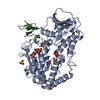
 Human herpesvirus 5
 X-RAY DIFFRACTION /
  Authors
 Citation
 Structure visualization
 Downloads & links
 Other downloads
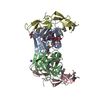


 PDBj
PDBj Assembly
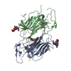

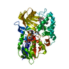
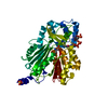
 Spodoptera frugiperda (fall armyworm) / References: UniProt: Q6RJQ3
 Sample preparation
 / Beamline: BL9-2 / Wavelength: 0.9795
  Processing