 Movie
Movie Controller


+ Open data

- Basic information
| Entry | Database: PDB / ID: 4jle | ||||||
|---|---|---|---|---|---|---|---|
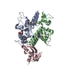
| Title | Structure of the P. falciparum PFI1780w PHIST domain | ||||||
 Components Components | PHIST | ||||||
 Keywords Keywords | PROTEIN BINDING / 3-helix bundle / Protein interaction module / PfEMP1 ATS domain / Human P. falciparum-infected erythrocytes | ||||||
| Function / homology |  Function and homology information Function and homology informationMaurer's cleft / adhesion of symbiont to microvasculature / host cell cytosol / host cell cytoplasm / host cell plasma membrane / membrane Similarity search - Function | ||||||
| Biological species |  | ||||||
| Method |  X-RAY DIFFRACTION / SYNCHROTRON / SAD / Resolution: 2.35 Å X-RAY DIFFRACTION / SYNCHROTRON / SAD / Resolution: 2.35 Å | ||||||
 Authors Authors | Slater, L. / Vakonakis, I. | ||||||
 Citation Citation | Journal: Faseb J. / Year: 2014 Title: A Plasmodium falciparum PHIST protein binds the virulence factor PfEMP1 and comigrates to knobs on the host cell surface. Authors: Oberli, A. / Slater, L.M. / Cutts, E. / Brand, F. / Mundwiler-Pachlatko, E. / Rusch, S. / Masik, M.F. / Erat, M.C. / Beck, H.P. / Vakonakis, I. | ||||||
| History |
|
- Structure visualization
| Structure viewer | Molecule: MolmilJmol/JSmol |
|---|
- Downloads & links
-Download
| PDBx/mmCIF format | 4jle.cif.gz | 146 KB | Display | PDBx/mmCIF format |
|---|---|---|---|---|
| PDB format | pdb4jle.ent.gz | 117.1 KB | Display | PDB format |
| PDBx/mmJSON format | 4jle.json.gz | Tree view | PDBx/mmJSON format | |
| Others |  Other downloads Other downloads |
-Validation report
| Arichive directory | https://data.pdbj.org/pub/pdb/validation_reports/jl/4jleftp://data.pdbj.org/pub/pdb/validation_reports/jl/4jle | HTTPS FTP |
|---|
-Related structure data
| Similar structure data |
|---|
-Links
 PDBj
PDBj- Assembly
| Deposited unit | 
| ||||||||
|---|---|---|---|---|---|---|---|---|---|
| 1 |
| ||||||||
| 2 | 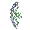
| ||||||||
| 3 | 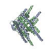
| ||||||||
| Unit cell |
| ||||||||
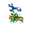
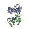
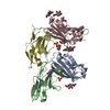
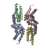
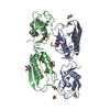
| Details | The asymmetric unit contains one helix-swapped protein dimer, however in solution under physiological conditions the protein is a monomer |
-Components
| #1: Protein | Mass: 20518.676 Da / Num. of mol.: 2 Source method: isolated from a genetically manipulated source Source: (gene. exp.) Strain: P. falciparum 3D7 / Gene: PFI1780w / Plasmid: PGEX-6P-2 / Production host:  #2: Chemical | ChemComp-CL /   Mass: 35.453 Da / Num. of mol.: 6 / Source method: obtained synthetically / Formula: Cl Mass: 35.453 Da / Num. of mol.: 6 / Source method: obtained synthetically / Formula: Cl#3: Chemical | ChemComp-ACT / |   Mass: 59.044 Da / Num. of mol.: 1 / Source method: obtained synthetically / Formula: C2H3O2 Mass: 59.044 Da / Num. of mol.: 1 / Source method: obtained synthetically / Formula: C2H3O2#4: Water | ChemComp-HOH / |  Mass: 18.015 Da / Num. of mol.: 47 / Source method: isolated from a natural source / Formula: H2O Mass: 18.015 Da / Num. of mol.: 47 / Source method: isolated from a natural source / Formula: H2O |
|---|
-Experimental details
-Experiment
| Experiment | Method: X-RAY DIFFRACTION / Number of used crystals: 1 |
|---|
- Sample preparation
| Crystal | Density Matthews: 2.6 Å3/Da / Density % sol: 52.65 % |
|---|---|
| Crystal grow | Temperature: 298 K / Method: vapor diffusion, sitting drop / pH: 4.6 Details: 2.0M Sodium Chloride 0.1M Sodium Acetate 22.5% Glycerol, pH 4.6, VAPOR DIFFUSION, SITTING DROP, temperature 298K |
-Data collection
| Diffraction | Mean temperature: 100 K |
|---|---|
| Diffraction source | Source: SYNCHROTRON / Site: Diamond  / Beamline: I04 / Wavelength: 0.9795 Å / Beamline: I04 / Wavelength: 0.9795 Å |
| Detector | Type: ADSC QUANTUM 315r / Detector: CCD / Date: Dec 10, 2011 |
| Radiation | Monochromator: Double crystal monochromator / Protocol: SINGLE WAVELENGTH / Monochromatic (M) / Laue (L): M / Scattering type: x-ray |
| Radiation wavelength | Wavelength: 0.9795 Å / Relative weight: 1 |
| Reflection | Resolution: 2.35→146.4 Å / Num. all: 18438 / Num. obs: 18438 / % possible obs: 99.9 % / Observed criterion σ(F): 0 / Observed criterion σ(I): 0 / Redundancy: 3.6 % / Biso Wilson estimate: 53.38 Å2 / Rmerge(I) obs: 0.063 / Rsym value: 0.063 / Net I/σ(I): 11.8 |
| Reflection shell | Resolution: 2.35→2.48 Å / Redundancy: 3.7 % / Rmerge(I) obs: 0.494 / Mean I/σ(I) obs: 2.4 / Num. unique all: 2646 / Rsym value: 0.494 / % possible all: 100 |
- Processing
| Software | Name: BUSTER / Version: 2.10.0 / Classification: refinement | |||||||||||||||||||||||||||||||||||||||||||||||||||||||||||||||||||||||||||
|---|---|---|---|---|---|---|---|---|---|---|---|---|---|---|---|---|---|---|---|---|---|---|---|---|---|---|---|---|---|---|---|---|---|---|---|---|---|---|---|---|---|---|---|---|---|---|---|---|---|---|---|---|---|---|---|---|---|---|---|---|---|---|---|---|---|---|---|---|---|---|---|---|---|---|---|---|
| Refinement | Method to determine structure: SAD / Resolution: 2.35→56.71 Å / Cor.coef. Fo:Fc: 0.9337 / Cor.coef. Fo:Fc free: 0.8976 / SU R Cruickshank DPI: 0.295 Isotropic thermal model: Isotropic thermal model with 1 TLS group per chain Cross valid method: THROUGHOUT / σ(F): 0 / Stereochemistry target values: Engh & Huber
| |||||||||||||||||||||||||||||||||||||||||||||||||||||||||||||||||||||||||||
| Displacement parameters | Biso mean: 66.53 Å2
| |||||||||||||||||||||||||||||||||||||||||||||||||||||||||||||||||||||||||||
| Refine analyze | Luzzati coordinate error obs: 0.369 Å | |||||||||||||||||||||||||||||||||||||||||||||||||||||||||||||||||||||||||||
| Refinement step | Cycle: LAST / Resolution: 2.35→56.71 Å
| |||||||||||||||||||||||||||||||||||||||||||||||||||||||||||||||||||||||||||
| Refine LS restraints |
| |||||||||||||||||||||||||||||||||||||||||||||||||||||||||||||||||||||||||||
| LS refinement shell | Resolution: 2.35→2.49 Å / Total num. of bins used: 9
| |||||||||||||||||||||||||||||||||||||||||||||||||||||||||||||||||||||||||||
| Refinement TLS params. | Method: refined / Refine-ID: X-RAY DIFFRACTION
| |||||||||||||||||||||||||||||||||||||||||||||||||||||||||||||||||||||||||||
| Refinement TLS group |
|
