- PDB-4jhs: Crystal structure of a C-terminal two domain fragment of human be... -
+
Open data
ID or keywords:
Loading...
-
Basic information
Entry
Database: PDB / ID: 4jhs
Title
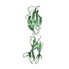
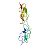
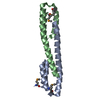
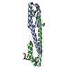
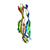
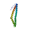
Crystal structure of a C-terminal two domain fragment of human beta-2-glycoprotein 1
Components
Beta-2-glycoprotein 1
Keywords
SIGNALING PROTEIN / STRUCTURAL GENOMICS / GLYCOPROTEIN / PSI-Biology / New York Structural Genomics Research Consortium / NYSGRC / Atoms-to-Animals: The Immune Function Network / IFN
Function / homology
Function and homology information
positive regulation of triglyceride metabolic process / lipoprotein lipase activator activity / triglyceride transport / chylomicron remodeling / platelet dense granule lumen / lipase binding / blood coagulation, intrinsic pathway / very-low-density lipoprotein particle remodeling / negative regulation of myeloid cell apoptotic process / regulation of fibrinolysis ...positive regulation of triglyceride metabolic process / lipoprotein lipase activator activity / triglyceride transport / chylomicron remodeling / platelet dense granule lumen / lipase binding / blood coagulation, intrinsic pathway / very-low-density lipoprotein particle remodeling / negative regulation of myeloid cell apoptotic process / regulation of fibrinolysis / chylomicron / high-density lipoprotein particle / very-low-density lipoprotein particle / negative regulation of endothelial cell migration / plasminogen activation / negative regulation of endothelial cell proliferation / negative regulation of smooth muscle cell apoptotic process / negative regulation of blood coagulation / negative regulation of fibrinolysis / positive regulation of blood coagulation / negative regulation of angiogenesis / phospholipid binding / Platelet degranulation / heparin binding / lipid binding / cell surface / : / extracellular exosome / extracellular region / identical protein binding Similarity search - Function
In the structure databanks used in Yorodumi, some data are registered as the other names, "COVID-19 virus" and "2019-nCoV". Here are the details of the virus and the list of structure data.
Jan 31, 2019. EMDB accession codes are about to change! (news from PDBe EMDB page)
EMDB accession codes are about to change! (news from PDBe EMDB page)
The allocation of 4 digits for EMDB accession codes will soon come to an end. Whilst these codes will remain in use, new EMDB accession codes will include an additional digit and will expand incrementally as the available range of codes is exhausted. The current 4-digit format prefixed with “EMD-” (i.e. EMD-XXXX) will advance to a 5-digit format (i.e. EMD-XXXXX), and so on. It is currently estimated that the 4-digit codes will be depleted around Spring 2019, at which point the 5-digit format will come into force.
The EM Navigator/Yorodumi systems omit the EMD- prefix.
Related info.:Q: What is EMD? / ID/Accession-code notation in Yorodumi/EM Navigator
Yorodumi is a browser for structure data from EMDB, PDB, SASBDB, etc.
This page is also the successor to EM Navigator detail page, and also detail information page/front-end page for Omokage search.
The word "yorodu" (or yorozu) is an old Japanese word meaning "ten thousand". "mi" (miru) is to see.
Related info.:EMDB / PDB / SASBDB / Comparison of 3 databanks / Yorodumi Search / Aug 31, 2016. New EM Navigator & Yorodumi / Yorodumi Papers / Jmol/JSmol / Function and homology information / Changes in new EM Navigator and Yorodumi
 Movie
Movie Controller

 Yorodumi
 Open data
 Basic information
 Components
 Keywords
 Function and homology information
 Homo sapiens (human)
 X-RAY DIFFRACTION /
  Authors
 Citation
 Structure visualization
 Downloads & links
 Other downloads


 PDBj
PDBj

 Assembly
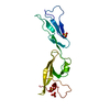
 TRICHOPLUSIA NI (cabbage looper) / Strain (production host): BTI-TN-5B1-4 / References: UniProt: P02749
 Mass: 96.063 Da / Num. of mol.: 3 / Source method: obtained synthetically / Formula: SO4
 Sample preparation
 / Beamline: X29A / Wavelength: 0.9795 Å
 Processing