- PDB-2g42: Crystal structure of a putative nucleic acid binding protein (tm0... -
+
Open data
ID or keywords:
Loading...
-
Basic information
Entry
Database: PDB / ID: 2g42
Title
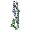
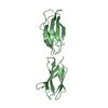
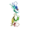
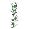
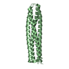
Crystal structure of a putative nucleic acid binding protein (tm0693) from thermotoga maritima at 2.28 A resolution
Components
hypothetical protein TM_0693
Keywords
GENE REGULATION / Structural genomics / Joint Center for Structural Genomics / JCSG / Protein Structure Initiative / PSI-2
Function / homology
Single alpha-helices involved in coiled-coils or other helix-helix interfaces - #50 / TM0693-like superfamily / Single alpha-helices involved in coiled-coils or other helix-helix interfaces / Helix non-globular / Special / metal ion binding / Flagellar protein FliT
Function and homology information
Biological species
Thermotoga maritima (bacteria)
Method
X-RAY DIFFRACTION / SYNCHROTRON / SAD / Resolution: 2.28 Å
BIOMOLECULE: 1 THIS ENTRY CONTAINS THE CRYSTALLOGRAPHIC ASYMMETRIC UNIT WHICH CONSISTS OF 2 CHAIN(S) ...BIOMOLECULE: 1 THIS ENTRY CONTAINS THE CRYSTALLOGRAPHIC ASYMMETRIC UNIT WHICH CONSISTS OF 2 CHAIN(S). SEE REMARK 350 FOR INFORMATION ON GENERATING THE BIOLOGICAL MOLECULE(S). SIZE EXCLUSION CHROMATOGRAPHY DATA SUPPORTS THE ASSIGNMENT OF A DIMER AS A BIOLOGICALLY SIGNIFICANT OLIGOMERIZATION STATE.
Remark 999
SEQUENCE THE CONSTRUCT WAS EXPRESSED WITH A PURIFICATION TAG MGSDKIHHHHHHENLYFQG. THE TAG WAS ...SEQUENCE THE CONSTRUCT WAS EXPRESSED WITH A PURIFICATION TAG MGSDKIHHHHHHENLYFQG. THE TAG WAS REMOVED WITH TEV PROTEASE LEAVING ONLY A GLYCINE (0) FOLLOWED BY THE TARGET SEQUENCE.
Mass: 18.015 Da / Num. of mol.: 72 / Source method: isolated from a natural source / Formula: H2O
Has protein modification
Y
-
Experimental details
-
Experiment
Experiment
Method: X-RAY DIFFRACTION / Number of used crystals: 1
-
Sample preparation
Crystal
Density Matthews: 2.18 Å3/Da / Density % sol: 43.23 % Description: AFTER THE INITIAL TRACE, A MODEL OF TM0693 IN A DIFFERENT CELL (PDB ENTRY 2FZT) WAS USED TO FACILITATE COMPLETION OF THE MODEL.
Crystal grow
Temperature: 277 K / Method: vapor diffusion, sitting drop, nanodrop / pH: 5.1 Details: 0.2M CaCl2, 20.0% PEG-3350, No Buffer, pH 5.1, VAPOR DIFFUSION, SITTING DROP, NANODROP, temperature 277K
Protocol: SINGLE WAVELENGTH / Monochromatic (M) / Laue (L): M / Scattering type: x-ray
Radiation wavelength
Wavelength: 0.9796 Å / Relative weight: 1
Reflection
Resolution: 2.28→40.859 Å / Num. obs: 7150 / % possible obs: 94.9 % / Redundancy: 6.777 % / Biso Wilson estimate: 42.374 Å2 / Rmerge(I) obs: 0.091 / Net I/σ(I): 12.72
Reflection shell
Diffraction-ID: 1
Resolution (Å)
Highest resolution (Å)
Redundancy (%)
Rmerge(I) obs
Mean I/σ(I) obs
Num. measured obs
Num. unique obs
% possible all
2.28-2.35
3.398
0.475
2.73
1334
385
66.4
2.35-2.44
0.452
4
3610
630
84.8
2.44-2.56
0.392
5
4814
742
94.4
2.56-2.69
0.324
6.4
5000
689
95.4
2.69-2.86
0.219
9.1
5461
734
97.1
2.86-3.08
0.158
11.6
5532
742
97.9
3.08-3.39
0.104
16.4
5523
746
98.5
3.39-3.87
0.073
21.3
5401
740
98.4
3.87
0.055
25.8
5557
766
98.7
-
Phasing
Phasing
Method: SAD
-
Processing
Software
Name
Version
Classification
NB
REFMAC
5.2.0005
refinement
XSCALE
datascaling
PDB_EXTRACT
1.701
dataextraction
XDS
datareduction
SOLVE
phasing
Refinement
Method to determine structure: SAD / Resolution: 2.28→40.8 Å / Cor.coef. Fo:Fc: 0.947 / Cor.coef. Fo:Fc free: 0.914 / SU B: 17.565 / SU ML: 0.22 / TLS residual ADP flag: LIKELY RESIDUAL / Cross valid method: THROUGHOUT / σ(F): 0 / ESU R: 0.438 / ESU R Free: 0.26 Stereochemistry target values: MAXIMUM LIKELIHOOD WITH PHASES Details: 1. HYDROGENS HAVE BEEN ADDED IN THE RIDING POSITIONS. 2. A MET-INHIBITION PROTOCOL WAS USED FOR SELENOMETHIONINE INCORPORATION DURING PROTEIN EXPRESSION. THE OCCUPANCY OF THE SE ATOMS IN THE ...Details: 1. HYDROGENS HAVE BEEN ADDED IN THE RIDING POSITIONS. 2. A MET-INHIBITION PROTOCOL WAS USED FOR SELENOMETHIONINE INCORPORATION DURING PROTEIN EXPRESSION. THE OCCUPANCY OF THE SE ATOMS IN THE MSE RESIDUES WAS REDUCED TO 0.75 TO ACCOUNT FOR THE REDUCED SCATTERING POWER DUE TO PARTIAL S-MET INCORPORATION.
Rfactor
Num. reflection
% reflection
Selection details
Rfree
0.251
330
4.6 %
RANDOM
Rwork
0.203
-
-
-
obs
0.206
7134
94.84 %
-
Solvent computation
Ion probe radii: 0.8 Å / Shrinkage radii: 0.8 Å / VDW probe radii: 1.2 Å / Solvent model: MASK
In the structure databanks used in Yorodumi, some data are registered as the other names, "COVID-19 virus" and "2019-nCoV". Here are the details of the virus and the list of structure data.
Jan 31, 2019. EMDB accession codes are about to change! (news from PDBe EMDB page)
EMDB accession codes are about to change! (news from PDBe EMDB page)
The allocation of 4 digits for EMDB accession codes will soon come to an end. Whilst these codes will remain in use, new EMDB accession codes will include an additional digit and will expand incrementally as the available range of codes is exhausted. The current 4-digit format prefixed with “EMD-” (i.e. EMD-XXXX) will advance to a 5-digit format (i.e. EMD-XXXXX), and so on. It is currently estimated that the 4-digit codes will be depleted around Spring 2019, at which point the 5-digit format will come into force.
The EM Navigator/Yorodumi systems omit the EMD- prefix.
Related info.:Q: What is EMD? / ID/Accession-code notation in Yorodumi/EM Navigator
Yorodumi is a browser for structure data from EMDB, PDB, SASBDB, etc.
This page is also the successor to EM Navigator detail page, and also detail information page/front-end page for Omokage search.
The word "yorodu" (or yorozu) is an old Japanese word meaning "ten thousand". "mi" (miru) is to see.
Related info.:EMDB / PDB / SASBDB / Comparison of 3 databanks / Yorodumi Search / Aug 31, 2016. New EM Navigator & Yorodumi / Yorodumi Papers / Jmol/JSmol / Function and homology information / Changes in new EM Navigator and Yorodumi
 Movie
Movie Controller

 Yorodumi
 Open data
 Basic information
 Components
 Keywords
 Function and homology information
 Thermotoga maritima (bacteria)
 X-RAY DIFFRACTION /
  Authors
 Citation
 Structure visualization
 Downloads & links
 Other downloads


 PDBj
PDBj Assembly
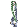

 Mass: 40.078 Da / Num. of mol.: 1 / Source method: obtained synthetically / Formula: Ca
 Mass: 18.015 Da / Num. of mol.: 72 / Source method: isolated from a natural source / Formula: H2O
 Sample preparation
 / Beamline: 8.2.1 / Wavelength: 0.9796
  Processing