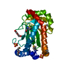
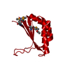
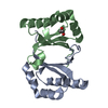
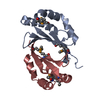
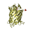
[English] 日本語
Yorodumi
- PDB-4jeg: Crystal Structure of Monobody CS1/SHP2 C-SH2 Domain Complex -

+
Open data


ID or keywords:

Loading...

-
Basic information

Entry
Database: PDB / ID: 4jeg
TitleCrystal Structure of Monobody CS1/SHP2 C-SH2 Domain Complex
Components
  • Monobody CS1
  • Tyrosine-protein phosphatase non-receptor type 11
KeywordsSignaling protein/protein binding / engineered binding protein / SHP2 SH2-monobody complex / Phosphatase / Phosphotyrosine Binding / Phosphorylation / Signaling protein-protein binding complex
Function / homology
Function and homology information


negative regulation of monocyte activation / calcium-independent cell-matrix adhesion / negative regulation of transforming growth factor beta production / Fibronectin matrix formation / negative regulation of cortisol secretion / intestinal epithelial cell migration / microvillus organization / negative regulation of growth hormone secretion / Extracellular matrix organization / positive regulation of substrate-dependent cell migration, cell attachment to substrate ...negative regulation of monocyte activation / calcium-independent cell-matrix adhesion / negative regulation of transforming growth factor beta production / Fibronectin matrix formation / negative regulation of cortisol secretion / intestinal epithelial cell migration / microvillus organization / negative regulation of growth hormone secretion / Extracellular matrix organization / positive regulation of substrate-dependent cell migration, cell attachment to substrate / genitalia development / neural crest cell migration involved in autonomic nervous system development / atrioventricular canal development / peptidase activator activity / fibrinogen complex / negative regulation of cell adhesion mediated by integrin / peptide cross-linking / STAT5 Activation / Netrin mediated repulsion signals / integrin activation / positive regulation of hormone secretion / cerebellar cortex formation / ALK mutants bind TKIs / multicellular organismal reproductive process / cell-substrate junction assembly / regulation of protein export from nucleus / positive regulation of ossification / hormone metabolic process / Interleukin-37 signaling / negative regulation of chondrocyte differentiation / Signaling by Leptin / MET activates PTPN11 / Regulation of RUNX1 Expression and Activity / face morphogenesis / biological process involved in interaction with symbiont / ERBB signaling pathway / Molecules associated with elastic fibres / Costimulation by the CD28 family / proteoglycan binding / Signal regulatory protein family interactions / peptide hormone receptor binding / triglyceride metabolic process / platelet formation / negative regulation of type I interferon production / extracellular matrix structural constituent / MET activates PTK2 signaling / megakaryocyte development / Syndecan interactions / organ growth / CTLA4 inhibitory signaling / Platelet sensitization by LDL / Interleukin-20 family signaling / PI-3K cascade:FGFR3 / Interleukin-6 signaling / p130Cas linkage to MAPK signaling for integrins / STAT5 activation downstream of FLT3 ITD mutants / PI-3K cascade:FGFR2 / PI-3K cascade:FGFR4 / Prolactin receptor signaling / PI-3K cascade:FGFR1 / endodermal cell differentiation / MAPK3 (ERK1) activation / phosphoprotein phosphatase activity / inner ear development / MAPK1 (ERK2) activation / PECAM1 interactions / Bergmann glial cell differentiation / regulation of cell adhesion mediated by integrin / neurotrophin TRK receptor signaling pathway / regulation of type I interferon-mediated signaling pathway / GRB2:SOS provides linkage to MAPK signaling for Integrins / platelet-derived growth factor receptor signaling pathway / non-membrane spanning protein tyrosine phosphatase activity / Signaling by ALK fusions and activated point mutants / Non-integrin membrane-ECM interactions / RET signaling / peptidyl-tyrosine dephosphorylation / basement membrane / Interleukin-3, Interleukin-5 and GM-CSF signaling / PI3K Cascade / Regulation of IFNA/IFNB signaling / endoplasmic reticulum-Golgi intermediate compartment / ECM proteoglycans / PD-1 signaling / negative regulation of insulin secretion / ephrin receptor signaling pathway / regulation of protein-containing complex assembly / fibroblast growth factor receptor signaling pathway / positive regulation of axon extension / GAB1 signalosome / Integrin cell surface interactions / Activated NTRK2 signals through FRS2 and FRS3 / Nuclear events stimulated by ALK signaling in cancer / Regulation of IFNG signaling / positive regulation of insulin receptor signaling pathway / homeostasis of number of cells within a tissue / FRS-mediated FGFR3 signaling / Signaling by CSF3 (G-CSF) / Signaling by FLT3 ITD and TKD mutants / FRS-mediated FGFR2 signaling
Similarity search - Function
Fibronectin type I domain / Fibronectin, type I / Fibronectin type-I domain signature. / Fibronectin type-I domain profile. / Fibronectin type 1 domain / Protein-tyrosine phosphatase, non-receptor type-6, -11 / Fibronectin type II domain / Fibronectin type II domain superfamily / Fibronectin type II domain / Fibronectin type-II collagen-binding domain signature. ...Fibronectin type I domain / Fibronectin, type I / Fibronectin type-I domain signature. / Fibronectin type-I domain profile. / Fibronectin type 1 domain / Protein-tyrosine phosphatase, non-receptor type-6, -11 / Fibronectin type II domain / Fibronectin type II domain superfamily / Fibronectin type II domain / Fibronectin type-II collagen-binding domain signature. / Fibronectin type-II collagen-binding domain profile. / Fibronectin type 2 domain / SH2 domain / SHC Adaptor Protein / Protein tyrosine phosphatase, catalytic domain / PTP type protein phosphatase domain profile. / Protein-tyrosine phosphatase / Tyrosine-specific protein phosphatase, PTPase domain / Protein-tyrosine phosphatase, catalytic / Protein tyrosine phosphatase, catalytic domain motif / Tyrosine specific protein phosphatases active site. / Protein-tyrosine phosphatase, active site / Kringle-like fold / Tyrosine-specific protein phosphatases domain / Tyrosine specific protein phosphatases domain profile. / Protein-tyrosine phosphatase-like / EGF-like domain signature 1. / Fibronectin type III domain / Fibronectin type 3 domain / Fibronectin type-III domain profile. / Fibronectin type III / Fibronectin type III superfamily / SH2 domain / Src homology 2 (SH2) domain profile. / Src homology 2 domains / SH2 domain / SH2 domain superfamily / Immunoglobulins / Immunoglobulin-like fold / Immunoglobulin-like / Sandwich / 2-Layer Sandwich / Mainly Beta / Alpha Beta
Similarity search - Domain/homology
Fibronectin / Tyrosine-protein phosphatase non-receptor type 11
Similarity search - Component
Biological speciesHomo sapiens (human)
MethodX-RAY DIFFRACTION / SYNCHROTRON / MOLECULAR REPLACEMENT / molecular replacement / Resolution: 2.3 Å
AuthorsSha, F. / Koide, S.
CitationJournal: Proc.Natl.Acad.Sci.USA / Year: 2013
Title: Dissection of the BCR-ABL signaling network using highly specific monobody inhibitors to the SHP2 SH2 domains.
Authors: Sha, F. / Gencer, E.B. / Georgeon, S. / Koide, A. / Yasui, N. / Koide, S. / Hantschel, O.
History
DepositionFeb 26, 2013Deposition site: RCSB / Processing site: RCSB
Revision 1.0Aug 28, 2013Provider: repository / Type: Initial release
Revision 1.1Sep 11, 2013Group: Database references
Revision 1.2Sep 25, 2013Group: Database references
Revision 1.3Nov 20, 2013Group: Data collection
Revision 1.4Mar 12, 2014Group: Structure summary

-
Structure visualization

Structure viewerMolecule:
MolmilJmol/JSmol

Downloads & links

-
Assembly

Deposited unit
A: Tyrosine-protein phosphatase non-receptor type 11
B: Monobody CS1


Theoretical massNumber of molelcules
Total (without water)24,4772
Polymers24,4772
Non-polymers00
Water1,44180
1


  • Idetical with deposited unit
  • defined by author&software
TypeNameSymmetry operationNumber
identity operation1_555x,y,z1
Buried area2110 Å2
ΔGint-11 kcal/mol
Surface area10480 Å2
MethodPISA
Unit cell
Length a, b, c (Å)111.700, 111.700, 31.780
Angle α, β, γ (deg.)90.000, 90.000, 120.000
Int Tables number169
Space group name H-MP61

-
Components

#1: Protein Tyrosine-protein phosphatase non-receptor type 11 / Protein-tyrosine phosphatase 1D / PTP-1D / Protein-tyrosine phosphatase 2C / PTP-2C / SH-PTP2 / SHP- ...Protein-tyrosine phosphatase 1D / PTP-1D / Protein-tyrosine phosphatase 2C / PTP-2C / SH-PTP2 / SHP-2 / Shp2 / SH-PTP3


Mass: 14006.806 Da / Num. of mol.: 1 / Fragment: C-terminal SH2 domain
Source method: isolated from a genetically manipulated source
Source: (gene. exp.) Homo sapiens (human) / Gene: PTP2C, PTPN11, SHPTP2 / Plasmid: pHBT / Production host: Escherichia coli (E. coli) / Strain (production host): BL21(DE3) / References: UniProt: Q06124
#2: Protein Monobody CS1


Mass: 10470.542 Da / Num. of mol.: 1
Source method: isolated from a genetically manipulated source
Source: (gene. exp.) Homo sapiens (human) / Plasmid: pHFT2 / Production host: Escherichia coli (E. coli) / Strain (production host): BL21(DE3) / References: UniProt: P02751*PLUS
#3: Water ChemComp-HOH / water / Water


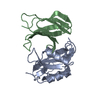
Mass: 18.015 Da / Num. of mol.: 80 / Source method: isolated from a natural source / Formula: H2O

-
Experimental details

-
Experiment

ExperimentMethod: X-RAY DIFFRACTION / Number of used crystals: 1

-
Sample preparation

CrystalDensity Matthews: 2.34 Å3/Da / Density % sol: 47.39 %
Crystal growTemperature: 293 K / Method: vapor diffusion, hanging drop / pH: 6.5
Details: 25% PEG 3350, 0.1M Bis-tris, 0.2M Sodium Chloride, pH 6.5, VAPOR DIFFUSION, HANGING DROP, temperature 293K

-
Data collection

DiffractionMean temperature: 100 K
Diffraction sourceSource: SYNCHROTRON / Site: APS / Beamline: 24-ID-E / Wavelength: 0.97918 Å
DetectorType: ADSC QUANTUM 315 / Detector: CCD / Date: Jun 20, 2012
RadiationMonochromator: Si(220) / Protocol: SINGLE WAVELENGTH / Monochromatic (M) / Laue (L): M / Scattering type: x-ray
Radiation wavelengthWavelength: 0.97918 Å / Relative weight: 1
ReflectionResolution: 2.3→19.35 Å / Num. all: 10400 / Num. obs: 10358 / % possible obs: 99.6 % / Observed criterion σ(F): 0 / Observed criterion σ(I): 3 / Redundancy: 7.9 % / Rmerge(I) obs: 0.062 / Net I/σ(I): 21.2
Reflection shellResolution: 2.3→2.42 Å / Redundancy: 7.4 % / Rmerge(I) obs: 0.423 / Mean I/σ(I) obs: 4.4 / Num. unique all: 1503 / % possible all: 99.6

-
Phasing

PhasingMethod: molecular replacement

-
Processing

Software
NameVersionClassificationNB
XSCALEdata scaling
PHASERphasing
PHENIX1.7.3_928refinement
PDB_EXTRACT3.11data extraction
ADSCQuantumdata collection
RefinementMethod to determine structure: MOLECULAR REPLACEMENT / Resolution: 2.3→19.347 Å / Occupancy max: 1 / Occupancy min: 1 / FOM work R set: 0.8461 / SU ML: 0.26 / σ(F): 1.44 / Phase error: 22.11 / Stereochemistry target values: ML
RfactorNum. reflection% reflectionSelection details
Rfree0.2268 498 4.81 %random
Rwork0.1868 ---
obs0.1888 10352 99.78 %-
all-10375 --
Solvent computationShrinkage radii: 0.6 Å / VDW probe radii: 0.9 Å / Solvent model: FLAT BULK SOLVENT MODEL / Bsol: 40.722 Å2 / ksol: 0.404 e/Å3
Displacement parametersBiso max: 103.27 Å2 / Biso mean: 34.5368 Å2 / Biso min: 10.39 Å2
Baniso -1Baniso -2Baniso -3
1--0.2239 Å20 Å2-0 Å2
2---0.2239 Å20 Å2
3---0.4479 Å2
Refinement stepCycle: LAST / Resolution: 2.3→19.347 Å
ProteinNucleic acidLigandSolventTotal
Num. atoms1638 0 0 80 1718
Refine LS restraints
Refine-IDTypeDev idealNumber
X-RAY DIFFRACTIONf_bond_d0.0041682
X-RAY DIFFRACTIONf_angle_d0.8852284
X-RAY DIFFRACTIONf_chiral_restr0.062249
X-RAY DIFFRACTIONf_plane_restr0.004287
X-RAY DIFFRACTIONf_dihedral_angle_d14.989605
LS refinement shell

Refine-ID: X-RAY DIFFRACTION / Total num. of bins used: 4

Resolution (Å)Rfactor RfreeNum. reflection RfreeRfactor RworkNum. reflection RworkNum. reflection all% reflection obs (%)
2.3-2.52990.31411150.2372420253599
2.5299-2.89490.30471320.215824412573100
2.8949-3.64330.20681300.177224422572100
3.6433-19.34770.18631210.168325512672100

+
About Yorodumi

-
News

-
Feb 9, 2022. New format data for meta-information of EMDB entries

New format data for meta-information of EMDB entries

  • Version 3 of the EMDB header file is now the official format.
  • The previous official version 1.9 will be removed from the archive.

Related info.:EMDB header

External links:wwPDB to switch to version 3 of the EMDB data model

-
Aug 12, 2020. Covid-19 info

Covid-19 info

URL: https://pdbj.org/emnavi/covid19.php

New page: Covid-19 featured information page in EM Navigator.

Related info.:Covid-19 info / Mar 5, 2020. Novel coronavirus structure data

+
Mar 5, 2020. Novel coronavirus structure data

Novel coronavirus structure data

Related info.:Yorodumi Speices / Aug 12, 2020. Covid-19 info

External links:COVID-19 featured content - PDBj / Molecule of the Month (242):Coronavirus Proteases

+
Jan 31, 2019. EMDB accession codes are about to change! (news from PDBe EMDB page)

EMDB accession codes are about to change! (news from PDBe EMDB page)

  • The allocation of 4 digits for EMDB accession codes will soon come to an end. Whilst these codes will remain in use, new EMDB accession codes will include an additional digit and will expand incrementally as the available range of codes is exhausted. The current 4-digit format prefixed with “EMD-” (i.e. EMD-XXXX) will advance to a 5-digit format (i.e. EMD-XXXXX), and so on. It is currently estimated that the 4-digit codes will be depleted around Spring 2019, at which point the 5-digit format will come into force.
  • The EM Navigator/Yorodumi systems omit the EMD- prefix.

Related info.:Q: What is EMD? / ID/Accession-code notation in Yorodumi/EM Navigator

External links:EMDB Accession Codes are Changing Soon! / Contact to PDBj

+
Jul 12, 2017. Major update of PDB

Major update of PDB

  • wwPDB released updated PDB data conforming to the new PDBx/mmCIF dictionary.
  • This is a major update changing the version number from 4 to 5, and with Remediation, in which all the entries are updated.
  • In this update, many items about electron microscopy experimental information are reorganized (e.g. em_software).
  • Now, EM Navigator and Yorodumi are based on the updated data.

External links:wwPDB Remediation / Enriched Model Files Conforming to OneDep Data Standards Now Available in the PDB FTP Archive

-
Yorodumi

Thousand views of thousand structures

  • Yorodumi is a browser for structure data from EMDB, PDB, SASBDB, etc.
  • This page is also the successor to EM Navigator detail page, and also detail information page/front-end page for Omokage search.
  • The word "yorodu" (or yorozu) is an old Japanese word meaning "ten thousand". "mi" (miru) is to see.

Related info.:EMDB / PDB / SASBDB / Comparison of 3 databanks / Yorodumi Search / Aug 31, 2016. New EM Navigator & Yorodumi / Yorodumi Papers / Jmol/JSmol / Function and homology information / Changes in new EM Navigator and Yorodumi

Read more