 ムービー
ムービー コントローラー


+ データを開く

- 基本情報
基本情報
| 登録情報 | データベース: PDB / ID: 4j9c | ||||||
|---|---|---|---|---|---|---|---|
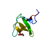
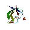
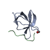
| タイトル | Crystal structure of the Abl-SH3 domain H59Q-N96T mutant complexed with the designed high-affinity peptide ligand P17 | ||||||
 要素 要素 |
| ||||||
 キーワード キーワード | Transferase/unknown function / beta shandwich / SH3 domain / kinase / poly proline rich motifs / Transferase-unknown function complex | ||||||
| 機能・相同性 |  機能・相同性情報 機能・相同性情報negative regulation of ubiquitin-protein transferase activity / protein localization to cytoplasmic microtubule plus-end / DNA conformation change / DN4 thymocyte differentiation / response to epinephrine / phospholipase C-inhibiting G protein-coupled receptor signaling pathway / podocyte apoptotic process / transitional one stage B cell differentiation / regulation of postsynaptic specialization assembly / positive regulation of phospholipase C/protein kinase C signal transduction ...negative regulation of ubiquitin-protein transferase activity / protein localization to cytoplasmic microtubule plus-end / DNA conformation change / DN4 thymocyte differentiation / response to epinephrine / phospholipase C-inhibiting G protein-coupled receptor signaling pathway / podocyte apoptotic process / transitional one stage B cell differentiation / regulation of postsynaptic specialization assembly / positive regulation of phospholipase C/protein kinase C signal transduction / regulation of modification of synaptic structure / nicotinate-nucleotide adenylyltransferase activity / delta-catenin binding / cerebellum morphogenesis / Role of ABL in ROBO-SLIT signaling / B cell proliferation involved in immune response / neuroepithelial cell differentiation / positive regulation of extracellular matrix organization / positive regulation of Wnt signaling pathway, planar cell polarity pathway / microspike assembly / B-1 B cell homeostasis / neuropilin signaling pathway / neuropilin binding / mitochondrial depolarization / regulation of cell motility / bubble DNA binding / positive regulation of establishment of T cell polarity / cellular response to dopamine / activated T cell proliferation / positive regulation of blood vessel branching / proline-rich region binding / negative regulation of mitotic cell cycle / regulation of Cdc42 protein signal transduction / mitogen-activated protein kinase binding / positive regulation of dendrite development / regulation of hematopoietic stem cell differentiation / syntaxin binding / alpha-beta T cell differentiation / regulation of axon extension / regulation of T cell differentiation / positive regulation of cell migration involved in sprouting angiogenesis / negative regulation of cell-cell adhesion / neuromuscular process controlling balance / Myogenesis / HDR through Single Strand Annealing (SSA) / positive regulation of osteoblast proliferation / platelet-derived growth factor receptor-beta signaling pathway / RUNX2 regulates osteoblast differentiation / Fc-gamma receptor signaling pathway involved in phagocytosis / vascular endothelial cell response to oscillatory fluid shear stress / Bergmann glial cell differentiation / regulation of endocytosis / negative regulation of cellular senescence / myoblast proliferation / negative regulation of long-term synaptic potentiation / regulation of microtubule polymerization / associative learning / positive regulation of vasoconstriction / actin monomer binding / positive regulation of focal adhesion assembly / negative regulation of BMP signaling pathway / ephrin receptor signaling pathway / RHO GTPases Activate WASPs and WAVEs / cardiac muscle cell proliferation / BMP signaling pathway / negative regulation of endothelial cell apoptotic process / cellular response to transforming growth factor beta stimulus / endothelial cell migration / positive regulation of T cell migration / negative regulation of double-strand break repair via homologous recombination / regulation of cell adhesion / mismatch repair / positive regulation of interleukin-2 production / ephrin receptor binding / spleen development / four-way junction DNA binding / ERK1 and ERK2 cascade / ruffle / positive regulation of stress fiber assembly / signal transduction in response to DNA damage / phosphotyrosine residue binding / actin filament polymerization / canonical NF-kappaB signal transduction / positive regulation of substrate adhesion-dependent cell spreading / substrate adhesion-dependent cell spreading / positive regulation of mitotic cell cycle / positive regulation of endothelial cell migration / SH2 domain binding / establishment of localization in cell / response to endoplasmic reticulum stress / Turbulent (oscillatory, disturbed) flow shear stress activates signaling by PIEZO1 and integrins in endothelial cells / thymus development / protein kinase C binding / positive regulation of release of sequestered calcium ion into cytosol / protein modification process / B cell receptor signaling pathway / regulation of autophagy / integrin-mediated signaling pathway / protein serine/threonine kinase activator activity / post-embryonic development 類似検索 - 分子機能 | ||||||
| 生物種 |  Homo sapiens (ヒト) Homo sapiens (ヒト) | ||||||
| 手法 |  X線回折 / シンクロトロン / 分子置換 / 解像度: 1.051 Å X線回折 / シンクロトロン / 分子置換 / 解像度: 1.051 Å | ||||||
 データ登録者 データ登録者 | Camara-Artigas, A. | ||||||
 引用 引用 | ジャーナル: To be Published タイトル: Crystal structure of the Abl-SH3 domain H59Q-N96T mutant complexed with the designed high-affinity peptide ligand P17 著者: Camara-Artigas, A. #1: ジャーナル: Acta Crystallogr.,Sect.D / 年: 2007タイトル: Crystallization by capillary counter-diffusion and structure determination of the N114A mutant of the SH3 domain of Abl tyrosine kinase complexed with a high-affinity peptide ligand. 著者: Camara-Artigas, A. / Palencia, A. / Martinez, J.C. / Luque, I. / Gavira, J.A. / Garcia-Ruiz, J.M. #2: ジャーナル: J.Biol.Chem. / 年: 2010タイトル: Role of interfacial water molecules in proline-rich ligand recognition by the Src homology 3 domain of Abl. 著者: Palencia, A. / Camara-Artigas, A. / Pisabarro, M.T. / Martinez, J.C. / Luque, I. | ||||||
| 履歴 |
|
- 構造の表示
構造の表示
| 構造ビューア | 分子: MolmilJmol/JSmol |
|---|
- ダウンロードとリンク
-ダウンロード
| PDBx/mmCIF形式 | 4j9c.cif.gz | 55.8 KB | 表示 | PDBx/mmCIF形式 |
|---|---|---|---|---|
| PDB形式 | pdb4j9c.ent.gz | 40.1 KB | 表示 | PDB形式 |
| PDBx/mmJSON形式 | 4j9c.json.gz | ツリー表示 | PDBx/mmJSON形式 | |
| その他 |  その他のダウンロード その他のダウンロード |
-検証レポート
| アーカイブディレクトリ | https://data.pdbj.org/pub/pdb/validation_reports/j9/4j9cftp://data.pdbj.org/pub/pdb/validation_reports/j9/4j9c | HTTPS FTP |
|---|
-関連構造データ
| 関連構造データ | 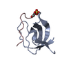 2o88S S: 精密化の開始モデル |
|---|---|
| 類似構造データ |


-リンク
 PDBj
PDBj


- 集合体
集合体
| 登録構造単位 | 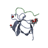
| ||||||||
|---|---|---|---|---|---|---|---|---|---|
| 1 |
| ||||||||
| 単位格子 |
| ||||||||
| Components on special symmetry positions |
|
-要素
| #1: タンパク質 | 分子量: 6986.677 Da / 分子数: 1 / 断片: SH3 domain (unp residues 60-121) / 変異: H59Q, N96T / 由来タイプ: 組換発現 / 由来: (組換発現) Homo sapiens (ヒト) / 遺伝子: ABL, ABL1, JTK7 / プラスミド: pET15b / 発現宿主:  参照: UniProt: P00519, non-specific protein-tyrosine kinase |
|---|---|
| #2: タンパク質・ペプチド | 分子量: 1065.218 Da / 分子数: 1 / 由来タイプ: 合成 |
| #3: 化合物 | ChemComp-PGE /   分子量: 150.173 Da / 分子数: 1 / 由来タイプ: 合成 / 式: C6H14O4 分子量: 150.173 Da / 分子数: 1 / 由来タイプ: 合成 / 式: C6H14O4 |
| #4: 化合物 | ChemComp-PEG /   分子量: 106.120 Da / 分子数: 1 / 由来タイプ: 合成 / 式: C4H10O3 分子量: 106.120 Da / 分子数: 1 / 由来タイプ: 合成 / 式: C4H10O3 |
| #5: 水 | ChemComp-HOH /  分子量: 18.015 Da / 分子数: 80 / 由来タイプ: 天然 / 式: H2O 分子量: 18.015 Da / 分子数: 80 / 由来タイプ: 天然 / 式: H2O |
| Has protein modification | Y |
-実験情報
-実験
| 実験 | 手法: X線回折 / 使用した結晶の数: 1 |
|---|
- 試料調製
試料調製
| 結晶 | マシュー密度: 2.01 Å3/Da / 溶媒含有率: 38.69 % |
|---|---|
| 結晶化 | 温度: 298 K / 手法: 蒸気拡散法, ハンギングドロップ法 / pH: 5.6 詳細: 1.5 M AMS, 5% PEG 200, 20 mM LiCl, 0.1 M AcONa , pH 5.6, VAPOR DIFFUSION, HANGING DROP, temperature 298K |
-データ収集
| 回折 | 平均測定温度: 100 K |
|---|---|
| 放射光源 | 由来: シンクロトロン / サイト: ESRF  / ビームライン: ID14-4 / 波長: 0.97 Å / ビームライン: ID14-4 / 波長: 0.97 Å |
| 検出器 | 検出器: CCD / 日付: 2011年4月8日 |
| 放射 | モノクロメーター: Channel cut ESRF monochromator and torodial focusing mirror プロトコル: SINGLE WAVELENGTH / 単色(M)・ラウエ(L): M / 散乱光タイプ: x-ray |
| 放射波長 | 波長: 0.97 Å / 相対比: 1 |
| 反射 | 解像度: 1.051→42.995 Å / Num. all: 29920 / Num. obs: 29561 / % possible obs: 98.8 % / Observed criterion σ(F): 0 / Observed criterion σ(I): 0 / 冗長度: 8.3 % / Biso Wilson estimate: 8.3 Å2 / Rmerge(I) obs: 0.04 / Rsym value: 0.04 / Net I/σ(I): 28.7 |
| 反射 シェル | 解像度: 1.05→1.11 Å / Rmerge(I) obs: 0.472 / Mean I/σ(I) obs: 4.61 / Num. unique all: 4721 / % possible all: 99.1 |
-位相決定
| 位相決定 | 手法: 分子置換 |
|---|
- 解析
解析
| ソフトウェア |
| ||||||||||||||||||||||||||||||||||||||||||||||||||||||||||||||||||||||||||||||||||||||||||||||||||||||||||||||||||||||||||||||||||||||||||||||||||||||||||
|---|---|---|---|---|---|---|---|---|---|---|---|---|---|---|---|---|---|---|---|---|---|---|---|---|---|---|---|---|---|---|---|---|---|---|---|---|---|---|---|---|---|---|---|---|---|---|---|---|---|---|---|---|---|---|---|---|---|---|---|---|---|---|---|---|---|---|---|---|---|---|---|---|---|---|---|---|---|---|---|---|---|---|---|---|---|---|---|---|---|---|---|---|---|---|---|---|---|---|---|---|---|---|---|---|---|---|---|---|---|---|---|---|---|---|---|---|---|---|---|---|---|---|---|---|---|---|---|---|---|---|---|---|---|---|---|---|---|---|---|---|---|---|---|---|---|---|---|---|---|---|---|---|---|---|---|
| 精密化 | 構造決定の手法: 分子置換 開始モデル: 2O88 解像度: 1.051→16.754 Å / Occupancy max: 1 / Occupancy min: 0 / SU ML: 0.08 / σ(F): 0 / σ(I): 0 / 位相誤差: 13.52 / 立体化学のターゲット値: ML
| ||||||||||||||||||||||||||||||||||||||||||||||||||||||||||||||||||||||||||||||||||||||||||||||||||||||||||||||||||||||||||||||||||||||||||||||||||||||||||
| 溶媒の処理 | 減衰半径: 0.9 Å / VDWプローブ半径: 1.11 Å / 溶媒モデル: FLAT BULK SOLVENT MODEL | ||||||||||||||||||||||||||||||||||||||||||||||||||||||||||||||||||||||||||||||||||||||||||||||||||||||||||||||||||||||||||||||||||||||||||||||||||||||||||
| 原子変位パラメータ | Biso max: 40.03 Å2 / Biso mean: 14.95 Å2 / Biso min: 6.31 Å2 | ||||||||||||||||||||||||||||||||||||||||||||||||||||||||||||||||||||||||||||||||||||||||||||||||||||||||||||||||||||||||||||||||||||||||||||||||||||||||||
| 精密化ステップ | サイクル: LAST / 解像度: 1.051→16.754 Å
| ||||||||||||||||||||||||||||||||||||||||||||||||||||||||||||||||||||||||||||||||||||||||||||||||||||||||||||||||||||||||||||||||||||||||||||||||||||||||||
| 拘束条件 |
| ||||||||||||||||||||||||||||||||||||||||||||||||||||||||||||||||||||||||||||||||||||||||||||||||||||||||||||||||||||||||||||||||||||||||||||||||||||||||||
| LS精密化 シェル | Refine-ID: X-RAY DIFFRACTION / Total num. of bins used: 21
|
