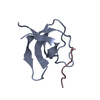
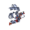
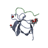
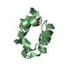
CD2-ASSOCIATEDPROTEIN / CAS LIGAND WITH MULTIPLE SH3 DOMAINS / ADAPTER PROTEIN CMS
Mass: 7412.285 Da / Num. of mol.: 1 / Fragment: SH3, RESIDUES 1-62 Source method: isolated from a genetically manipulated source Details: N-TERMINAL SH3 DOMAIN (SH3A) OF CD2- ASSOCIATED PROTEIN (CD2AP)OR CAS LIGAND WITH MULTIPLE SRC HOMOLOGY 3 DOMAINS (CMS) Source: (gene. exp.) HOMO SAPIENS (human) / Plasmid: PET21A / Production host: ESCHERICHIA COLI (E. coli) / Strain (production host): ROSETTA (DE3) PLYS / References: UniProt: Q9Y5K6
#2: Protein/peptide
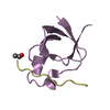
E3UBIQUITIN-PROTEINLIGASECBL-B / CAS-BR-M MURINE ECTROPIC RETROVIRAL TRANSFORMING SEQUENCE B / CBL-B / SIGNAL TRANSDUCTION PROTEIN ...CAS-BR-M MURINE ECTROPIC RETROVIRAL TRANSFORMING SEQUENCE B / CBL-B / SIGNAL TRANSDUCTION PROTEIN CBL-B / SH3-BINDING PROTEIN CBL-B / CASITAS B-LINEAGE LYMPHOMA PROTO-ONCOGENE B / RING FINGER PROTEIN 56
Mass: 1332.622 Da / Num. of mol.: 1 / Fragment: PEPTIDE, RESIDUES 902-912 / Source method: obtained synthetically Details: A.A. 902 TO 912 FROM CAS-BR-M (MURINE) ECTROPIC RETROVIRAL TRANSFORMING SEQUENCE B (CBL-B) Source: (synth.) HOMO SAPIENS (human) References: UniProt: Q13191, Ligases; Forming carbon-nitrogen bonds; Acid-amino-acid ligases (peptide synthases)
Protocol: SINGLE WAVELENGTH / Monochromatic (M) / Laue (L): M / Scattering type: x-ray
Radiation wavelength
Wavelength: 1.54179 Å / Relative weight: 1
Reflection
Resolution: 1.7→20 Å / Num. obs: 8678 / % possible obs: 99.9 % / Observed criterion σ(I): 3 / Redundancy: 12.33 % / Biso Wilson estimate: 33 Å2 / Rmerge(I) obs: 0.03 / Net I/σ(I): 20.9
Reflection shell
Resolution: 1.7→1.76 Å / Rmerge(I) obs: 0.27 / Mean I/σ(I) obs: 6.9 / % possible all: 100
-
Processing
Software
Name
Version
Classification
REFMAC
5.2.0019
refinement
DENZO
datareduction
SCALEPACK
datascaling
AMoRE
phasing
Refinement
Method to determine structure: MOLECULAR REPLACEMENT / Resolution: 1.7→19.32 Å / Cor.coef. Fo:Fc: 0.957 / Cor.coef. Fo:Fc free: 0.942 / SU B: 4.084 / SU ML: 0.07 / TLS residual ADP flag: LIKELY RESIDUAL / Cross valid method: THROUGHOUT / σ(F): 0 / ESU R: 0.112 / ESU R Free: 0.107 / Stereochemistry target values: MAXIMUM LIKELIHOOD Details: HYDROGENS HAVE BEEN ADDED IN THE RIDING POSITIONS.REFINENEMENT WAS INITIATED WITH CNS V1.1 AND PURSUED WITH REFMAC 5 DISORDERED ATOMS IN SIDE CHAIN S WHERE GIVEN AN OCCUPANCY CG LYS A 31, CD ...Details: HYDROGENS HAVE BEEN ADDED IN THE RIDING POSITIONS.REFINENEMENT WAS INITIATED WITH CNS V1.1 AND PURSUED WITH REFMAC 5 DISORDERED ATOMS IN SIDE CHAIN S WHERE GIVEN AN OCCUPANCY CG LYS A 31, CD LYS A 31, CE LYS A 31, NZ LYS A 31, CD GLU A 39, OE1 GLU A 39, OE2 GLU A 39, CE BMET A 48
Rfactor
Num. reflection
% reflection
Selection details
Rfree
0.218
465
5.44 %
RANDOM
Rwork
0.19
-
-
-
obs
0.192
8557
98.3 %
-
Solvent computation
Ion probe radii: 0.8 Å / Shrinkage radii: 0.8 Å / VDW probe radii: 1.4 Å / Solvent model: BABINET MODEL PLUS MASK
 Movie
Movie Controller

 Yorodumi
 Open data
 Basic information
 Components
 Keywords
 Function and homology information
 HOMO SAPIENS (human)
 X-RAY DIFFRACTION /
  Authors
 Citation
 Structure visualization
 Downloads & links
 Other downloads
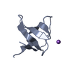


 PDBj
PDBj


 Assembly

 Mass: 18.015 Da / Num. of mol.: 60 / Source method: isolated from a natural source / Formula: H2O
 Sample preparation
 Processing