 Movie
Movie Controller

[English] 日本語
 Yorodumi
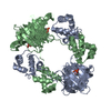
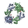
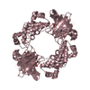
- PDB-4j1x: Crystal Structure of Fe(II)-HppE with alternative substrate (S)-1-HPP -
+ Open data

- Basic information
| Entry | Database: PDB / ID: 4j1x | ||||||
|---|---|---|---|---|---|---|---|
| Title | Crystal Structure of Fe(II)-HppE with alternative substrate (S)-1-HPP | ||||||
 Components Components | Epoxidase | ||||||
 Keywords Keywords | METAL BINDING PROTEIN / keto product / Hydroxypropylphosphonic acid epoxidase / mononuclear non-heme iron enzyme / cupin fold / phosphono migration | ||||||
| Function / homology |  Function and homology information Function and homology information(S)-2-hydroxypropylphosphonic acid epoxidase / phosphinothricin biosynthetic process / oxidoreductase activity, acting on paired donors, with oxidation of a pair of donors resulting in the reduction of molecular oxygen to two molecules of water / dioxygenase activity / antibiotic biosynthetic process / ferrous iron binding / protein homotetramerization / DNA-binding transcription factor activity / DNA binding / cytosol Similarity search - Function | ||||||
| Biological species |  Streptomyces wedmorensis (bacteria) Streptomyces wedmorensis (bacteria) | ||||||
| Method |  X-RAY DIFFRACTION / SYNCHROTRON / MOLECULAR REPLACEMENT / Resolution: 2.8 Å X-RAY DIFFRACTION / SYNCHROTRON / MOLECULAR REPLACEMENT / Resolution: 2.8 Å | ||||||
 Authors Authors | Drennan, C.L. / Dey, M. | ||||||
 Citation Citation | Journal: Nature / Year: 2013 Title: Mechanistic studies of an unprecedented enzyme-catalysed 1,2-phosphono-migration reaction. Authors: Chang, W.C. / Dey, M. / Liu, P. / Mansoorabadi, S.O. / Moon, S.J. / Zhao, Z.K. / Drennan, C.L. / Liu, H.W. | ||||||
| History |
|
- Structure visualization
| Structure viewer | Molecule: MolmilJmol/JSmol |
|---|
- Downloads & links
-Download
| PDBx/mmCIF format | 4j1x.cif.gz | 120.8 KB | Display | PDBx/mmCIF format |
|---|---|---|---|---|
| PDB format | pdb4j1x.ent.gz | 94.8 KB | Display | PDB format |
| PDBx/mmJSON format | 4j1x.json.gz | Tree view | PDBx/mmJSON format | |
| Others |  Other downloads Other downloads |
-Validation report
| Arichive directory | https://data.pdbj.org/pub/pdb/validation_reports/j1/4j1xftp://data.pdbj.org/pub/pdb/validation_reports/j1/4j1x | HTTPS FTP |
|---|
-Related structure data
| Related structure data |  4j1wC  1zz8S C: citing same article ( S: Starting model for refinement |
|---|---|
| Similar structure data |


-Links
 PDBj
PDBj


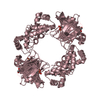
- Assembly
| Deposited unit | 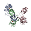
| ||||||||
|---|---|---|---|---|---|---|---|---|---|
| 1 | 
| ||||||||
| 2 | 
| ||||||||
| Unit cell |
|
-Components
| #1: Protein | Mass: 21229.932 Da / Num. of mol.: 3 Fragment: Metal and substrate binding domains (UNP Residues 2-198) Source method: isolated from a genetically manipulated source Source: (gene. exp.) Streptomyces wedmorensis (bacteria) / Gene: fom4 / Plasmid: pPL001 / Production host: #2: Chemical |   Mass: 55.845 Da / Num. of mol.: 3 / Source method: obtained synthetically / Formula: Fe Mass: 55.845 Da / Num. of mol.: 3 / Source method: obtained synthetically / Formula: Fe#3: Chemical | 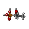  Mass: 140.075 Da / Num. of mol.: 3 / Source method: obtained synthetically / Formula: C3H9O4P Mass: 140.075 Da / Num. of mol.: 3 / Source method: obtained synthetically / Formula: C3H9O4P#4: Chemical | ChemComp-GOL / |   Mass: 92.094 Da / Num. of mol.: 1 / Source method: obtained synthetically / Formula: C3H8O3 Mass: 92.094 Da / Num. of mol.: 1 / Source method: obtained synthetically / Formula: C3H8O3#5: Water | ChemComp-HOH / |  Mass: 18.015 Da / Num. of mol.: 33 / Source method: isolated from a natural source / Formula: H2O Mass: 18.015 Da / Num. of mol.: 33 / Source method: isolated from a natural source / Formula: H2O |
|---|
-Experimental details
-Experiment
| Experiment | Method: X-RAY DIFFRACTION / Number of used crystals: 1 |
|---|
- Sample preparation
| Crystal | Density Matthews: 3.69 Å3/Da / Density % sol: 66.68 % |
|---|---|
| Crystal grow | Temperature: 298 K / Method: vapor diffusion, hanging drop / pH: 8.5 Details: 0.1 M Tris.HCl, pH 8.5, 2.0 M ammonium sulfate, VAPOR DIFFUSION, HANGING DROP, temperature 298K |
-Data collection
| Diffraction | Mean temperature: 100 K |
|---|---|
| Diffraction source | Source: SYNCHROTRON / Site: ALS  / Beamline: 8.2.2 / Wavelength: 1 Å / Beamline: 8.2.2 / Wavelength: 1 Å |
| Detector | Type: ADSC QUANTUM 315 / Detector: CCD / Date: Jul 30, 2010 |
| Radiation | Monochromator: Double crystal, Si(111) / Protocol: SINGLE WAVELENGTH / Monochromatic (M) / Laue (L): M / Scattering type: x-ray |
| Radiation wavelength | Wavelength: 1 Å / Relative weight: 1 |
| Reflection | Resolution: 2.8→50 Å / Num. obs: 23538 / % possible obs: 99.3 % / Observed criterion σ(F): 0 / Observed criterion σ(I): -3 |
| Reflection shell | Resolution: 2.8→2.85 Å / Redundancy: 8.7 % / Mean I/σ(I) obs: 2.7 / Rsym value: 0.741 / % possible all: 99.3 |
- Processing
| Software |
| ||||||||||||||||||||||||||||||||||||||||||||||||||||||||||||
|---|---|---|---|---|---|---|---|---|---|---|---|---|---|---|---|---|---|---|---|---|---|---|---|---|---|---|---|---|---|---|---|---|---|---|---|---|---|---|---|---|---|---|---|---|---|---|---|---|---|---|---|---|---|---|---|---|---|---|---|---|---|
| Refinement | Method to determine structure: MOLECULAR REPLACEMENT Starting model: PDB ENTRY 1ZZ8 Resolution: 2.8→50 Å / σ(F): 0 / Stereochemistry target values: Engh & Huber
| ||||||||||||||||||||||||||||||||||||||||||||||||||||||||||||
| Refinement step | Cycle: LAST / Resolution: 2.8→50 Å
| ||||||||||||||||||||||||||||||||||||||||||||||||||||||||||||
| Refine LS restraints |
| ||||||||||||||||||||||||||||||||||||||||||||||||||||||||||||
| LS refinement shell | Resolution: 2.8→2.85 Å |
