[English] 日本語
Yorodumi
- PDB-3scf: Fe(II)-HppE with S-HPP and NO -

+
Open data


ID or keywords:

Loading...

-
Basic information

Entry
Database: PDB / ID: 3scf
TitleFe(II)-HppE with S-HPP and NO
ComponentsEpoxidase
Keywords
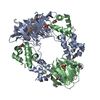
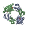
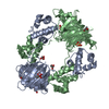
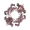
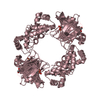
METAL BINDING PROTEIN / Cupin-fold / Hydroxypropylphosphonic acid epoxidase / mononuclear non-heme iron enzyme
Function / homology
Function and homology information


(S)-2-hydroxypropylphosphonic acid epoxidase / phosphinothricin biosynthetic process / oxidoreductase activity, acting on paired donors, with oxidation of a pair of donors resulting in the reduction of molecular oxygen to two molecules of water / dioxygenase activity / antibiotic biosynthetic process / ferrous iron binding / protein homotetramerization / DNA binding
Similarity search - Function
Cupin 2, conserved barrel / Cupin domain / Helix-turn-helix / Helix-turn-helix XRE-family like proteins / Cro/C1-type HTH domain profile. / lambda repressor-like DNA-binding domains / Cro/C1-type helix-turn-helix domain / 434 Repressor (Amino-terminal Domain) / Lambda repressor-like, DNA-binding domain superfamily / RmlC-like cupin domain superfamily ...Cupin 2, conserved barrel / Cupin domain / Helix-turn-helix / Helix-turn-helix XRE-family like proteins / Cro/C1-type HTH domain profile. / lambda repressor-like DNA-binding domains / Cro/C1-type helix-turn-helix domain / 434 Repressor (Amino-terminal Domain) / Lambda repressor-like, DNA-binding domain superfamily / RmlC-like cupin domain superfamily / Jelly Rolls / RmlC-like jelly roll fold / Jelly Rolls / Sandwich / Orthogonal Bundle / Mainly Beta / Mainly Alpha
Similarity search - Domain/homology
: / NITRIC OXIDE / (S)-2-HYDROXYPROPYLPHOSPHONIC ACID / (S)-2-hydroxypropylphosphonic acid epoxidase
Similarity search - Component
Biological speciesStreptomyces wedmorensis (bacteria)
MethodX-RAY DIFFRACTION / SYNCHROTRON / MOLECULAR REPLACEMENT / Resolution: 2.85 Å
AuthorsDrennan, C.L.
CitationJournal: J.Am.Chem.Soc. / Year: 2011
Title: Structural basis of regiospecificity of a mononuclear iron enzyme in antibiotic fosfomycin biosynthesis.
Authors: Yun, D. / Dey, M. / Higgins, L.J. / Yan, F. / Liu, H.W. / Drennan, C.L.
History
DepositionJun 7, 2011Deposition site: RCSB / Processing site: RCSB
Revision 1.0Jul 6, 2011Provider: repository / Type: Initial release
Revision 1.1Jul 13, 2011Group: Version format compliance
Revision 1.2Aug 10, 2011Group: Database references
Revision 1.3Sep 13, 2023Group: Data collection / Database references ...Data collection / Database references / Derived calculations / Refinement description
Category: chem_comp_atom / chem_comp_bond ...chem_comp_atom / chem_comp_bond / database_2 / pdbx_initial_refinement_model / pdbx_struct_conn_angle / struct_conn / struct_site
Item: _database_2.pdbx_DOI / _database_2.pdbx_database_accession ..._database_2.pdbx_DOI / _database_2.pdbx_database_accession / _pdbx_struct_conn_angle.ptnr1_auth_asym_id / _pdbx_struct_conn_angle.ptnr1_auth_comp_id / _pdbx_struct_conn_angle.ptnr1_auth_seq_id / _pdbx_struct_conn_angle.ptnr1_label_asym_id / _pdbx_struct_conn_angle.ptnr1_label_atom_id / _pdbx_struct_conn_angle.ptnr1_label_comp_id / _pdbx_struct_conn_angle.ptnr1_label_seq_id / _pdbx_struct_conn_angle.ptnr2_auth_asym_id / _pdbx_struct_conn_angle.ptnr2_auth_seq_id / _pdbx_struct_conn_angle.ptnr2_label_asym_id / _pdbx_struct_conn_angle.ptnr3_auth_asym_id / _pdbx_struct_conn_angle.ptnr3_auth_comp_id / _pdbx_struct_conn_angle.ptnr3_auth_seq_id / _pdbx_struct_conn_angle.ptnr3_label_asym_id / _pdbx_struct_conn_angle.ptnr3_label_atom_id / _pdbx_struct_conn_angle.ptnr3_label_comp_id / _pdbx_struct_conn_angle.ptnr3_label_seq_id / _pdbx_struct_conn_angle.value / _struct_conn.pdbx_dist_value / _struct_conn.ptnr1_auth_asym_id / _struct_conn.ptnr1_auth_comp_id / _struct_conn.ptnr1_auth_seq_id / _struct_conn.ptnr1_label_asym_id / _struct_conn.ptnr1_label_atom_id / _struct_conn.ptnr1_label_comp_id / _struct_conn.ptnr1_label_seq_id / _struct_conn.ptnr2_auth_asym_id / _struct_conn.ptnr2_auth_comp_id / _struct_conn.ptnr2_auth_seq_id / _struct_conn.ptnr2_label_asym_id / _struct_conn.ptnr2_label_atom_id / _struct_conn.ptnr2_label_comp_id / _struct_site.pdbx_auth_asym_id / _struct_site.pdbx_auth_comp_id / _struct_site.pdbx_auth_seq_id

-
Structure visualization

Structure viewerMolecule:
MolmilJmol/JSmol

Downloads & links

-
Assembly

Deposited unit
A: Epoxidase
B: Epoxidase
C: Epoxidase
hetero molecules


Theoretical massNumber of molelcules
Total (without water)64,91513
Polymers64,0833
Non-polymers83210
Water48627
1
A: Epoxidase
B: Epoxidase
hetero molecules

A: Epoxidase
B: Epoxidase
hetero molecules


Theoretical massNumber of molelcules
Total (without water)86,53218
Polymers85,4454
Non-polymers1,08814
Water724
TypeNameSymmetry operationNumber
identity operation1_555x,y,z1
crystal symmetry operation2_765-x+2,-y+1,z1
Buried area15650 Å2
ΔGint-156 kcal/mol
Surface area31220 Å2
MethodPISA
2
C: Epoxidase
hetero molecules

C: Epoxidase
hetero molecules

C: Epoxidase
hetero molecules

C: Epoxidase
hetero molecules


Theoretical massNumber of molelcules
Total (without water)86,59716
Polymers85,4454
Non-polymers1,15212
Water724
TypeNameSymmetry operationNumber
identity operation1_555x,y,z1
crystal symmetry operation2_665-x+1,-y+1,z1
crystal symmetry operation7_555y,x,-z1
crystal symmetry operation8_665-y+1,-x+1,-z1
Buried area16040 Å2
ΔGint-138 kcal/mol
Surface area30910 Å2
MethodPISA
Unit cell
Length a, b, c (Å)111.661, 111.661, 152.158
Angle α, β, γ (deg.)90.00, 90.00, 90.00
Int Tables number94
Space group name H-MP42212

-
Components

-
Protein , 1 types, 3 molecules ABC

#1: Protein Epoxidase


Mass: 21361.127 Da / Num. of mol.: 3
Source method: isolated from a genetically manipulated source
Source: (gene. exp.) Streptomyces wedmorensis (bacteria) / Gene: fom4 / Plasmid: pPL001 / Production host: Escherichia coli (E. coli) / Strain (production host): B834(DE3) / References: UniProt: Q56185

-
Non-polymers , 5 types, 37 molecules

#2: Chemical ChemComp-FE2 / FE (II) ION


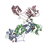
Mass: 55.845 Da / Num. of mol.: 3 / Source method: obtained synthetically / Formula: Fe
#3: Chemical ChemComp-NO / NITRIC OXIDE / Nitrogen monoxide


Mass: 30.006 Da / Num. of mol.: 2 / Source method: obtained synthetically / Formula: NO
#4: Chemical ChemComp-S0H / (S)-2-HYDROXYPROPYLPHOSPHONIC ACID


Mass: 140.075 Da / Num. of mol.: 3 / Source method: obtained synthetically / Formula: C3H9O4P
#5: Chemical ChemComp-GOL / GLYCEROL / GLYCERIN / PROPANE-1,2,3-TRIOL


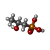
Mass: 92.094 Da / Num. of mol.: 2 / Source method: obtained synthetically / Formula: C3H8O3
#6: Water ChemComp-HOH / water


Mass: 18.015 Da / Num. of mol.: 27 / Source method: isolated from a natural source / Formula: H2O

-
Experimental details

-
Experiment

ExperimentMethod: X-RAY DIFFRACTION / Number of used crystals: 1

-
Sample preparation

CrystalDensity Matthews: 3.7 Å3/Da / Density % sol: 66.76 %
Crystal growTemperature: 298 K / Method: vapor diffusion, hanging drop / pH: 8.5
Details: 2.0 M ammonium sulfate, 0.1 M Tris-HCl, pH 8.5, VAPOR DIFFUSION, HANGING DROP, temperature 298K

-
Data collection

DiffractionMean temperature: 100 K
Diffraction sourceSource: SYNCHROTRON / Site: APS / Beamline: 24-ID-C / Wavelength: 0.97 Å
DetectorType: ADSC QUANTUM 315 / Detector: CCD / Date: Apr 26, 2007
RadiationMonochromator: Si(111) / Protocol: SINGLE WAVELENGTH / Monochromatic (M) / Laue (L): M / Scattering type: x-ray
Radiation wavelengthWavelength: 0.97 Å / Relative weight: 1
ReflectionResolution: 2.85→50 Å / Num. all: 22766 / Num. obs: 22766 / % possible obs: 99.1 % / Redundancy: 10.7 % / Rsym value: 0.101 / Net I/σ(I): 21.9
Reflection shellResolution: 2.85→2.95 Å / Mean I/σ(I) obs: 2.3 / Rsym value: 0.438 / % possible all: 92.8

-
Processing

Software
NameVersionClassification
ADSCQuantumdata collection
CNSrefinement
DENZOdata reduction
SCALEPACKdata scaling
CNSphasing
RefinementMethod to determine structure: MOLECULAR REPLACEMENT
Starting model: pdb entry 1ZZ8
Resolution: 2.85→50 Å / σ(F): 0 / Stereochemistry target values: Engh & Huber
RfactorNum. reflectionSelection details
Rfree0.269 1119 random
Rwork0.243 --
all0.269 22379 -
obs0.243 22379 -
Refinement stepCycle: LAST / Resolution: 2.85→50 Å
ProteinNucleic acidLigandSolventTotal
Num. atoms4357 0 43 27 4427
Refine LS restraints
Refine-IDTypeDev ideal
X-RAY DIFFRACTIONc_bond_d0.146
X-RAY DIFFRACTIONc_angle_d0.419

+
About Yorodumi

-
News

-
Feb 9, 2022. New format data for meta-information of EMDB entries

New format data for meta-information of EMDB entries

  • Version 3 of the EMDB header file is now the official format.
  • The previous official version 1.9 will be removed from the archive.

Related info.:EMDB header

External links:wwPDB to switch to version 3 of the EMDB data model

-
Aug 12, 2020. Covid-19 info

Covid-19 info

URL: https://pdbjlvh1.pdbj.org/emnavi/covid19.php

New page: Covid-19 featured information page in EM Navigator.

Related info.:Covid-19 info / Mar 5, 2020. Novel coronavirus structure data

+
Mar 5, 2020. Novel coronavirus structure data

Novel coronavirus structure data

Related info.:Yorodumi Speices / Aug 12, 2020. Covid-19 info

External links:COVID-19 featured content - PDBj / Molecule of the Month (242):Coronavirus Proteases

+
Jan 31, 2019. EMDB accession codes are about to change! (news from PDBe EMDB page)

EMDB accession codes are about to change! (news from PDBe EMDB page)

  • The allocation of 4 digits for EMDB accession codes will soon come to an end. Whilst these codes will remain in use, new EMDB accession codes will include an additional digit and will expand incrementally as the available range of codes is exhausted. The current 4-digit format prefixed with “EMD-” (i.e. EMD-XXXX) will advance to a 5-digit format (i.e. EMD-XXXXX), and so on. It is currently estimated that the 4-digit codes will be depleted around Spring 2019, at which point the 5-digit format will come into force.
  • The EM Navigator/Yorodumi systems omit the EMD- prefix.

Related info.:Q: What is EMD? / ID/Accession-code notation in Yorodumi/EM Navigator

External links:EMDB Accession Codes are Changing Soon! / Contact to PDBj

+
Jul 12, 2017. Major update of PDB

Major update of PDB

  • wwPDB released updated PDB data conforming to the new PDBx/mmCIF dictionary.
  • This is a major update changing the version number from 4 to 5, and with Remediation, in which all the entries are updated.
  • In this update, many items about electron microscopy experimental information are reorganized (e.g. em_software).
  • Now, EM Navigator and Yorodumi are based on the updated data.

External links:wwPDB Remediation / Enriched Model Files Conforming to OneDep Data Standards Now Available in the PDB FTP Archive

-
Yorodumi

Thousand views of thousand structures

  • Yorodumi is a browser for structure data from EMDB, PDB, SASBDB, etc.
  • This page is also the successor to EM Navigator detail page, and also detail information page/front-end page for Omokage search.
  • The word "yorodu" (or yorozu) is an old Japanese word meaning "ten thousand". "mi" (miru) is to see.

Related info.:EMDB / PDB / SASBDB / Comparison of 3 databanks / Yorodumi Search / Aug 31, 2016. New EM Navigator & Yorodumi / Yorodumi Papers / Jmol/JSmol / Function and homology information / Changes in new EM Navigator and Yorodumi

Read more