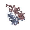
[English] 日本語
Yorodumi
- PDB-4itr: Crystal Structure of IbpAFic2-H3717A in complex with adenylylated... -

+
Open data


ID or keywords:

Loading...

-
Basic information

Entry
Database: PDB / ID: 4itr
TitleCrystal Structure of IbpAFic2-H3717A in complex with adenylylated Cdc42
Components
  • Adenosine monophosphate-protein transferase and cysteine protease IbpA
  • Cell division control protein 42 homolog
KeywordsTRANSFERASE / Fic domain / Adenosine monophosphate-protein transferase
Function / homology
Function and homology information


GBD domain binding / submandibular salivary gland formation / actin filament branching / Golgi transport complex / AMPylase activity / positive regulation of pinocytosis / modification of synaptic structure / protein adenylyltransferase / protein adenylylation / endothelin receptor signaling pathway involved in heart process ...GBD domain binding / submandibular salivary gland formation / actin filament branching / Golgi transport complex / AMPylase activity / positive regulation of pinocytosis / modification of synaptic structure / protein adenylyltransferase / protein adenylylation / endothelin receptor signaling pathway involved in heart process / Cdc42 protein signal transduction / cardiac neural crest cell migration involved in outflow tract morphogenesis / positive regulation of synapse structural plasticity / dendritic cell migration / storage vacuole / positive regulation of epithelial cell proliferation involved in lung morphogenesis / apolipoprotein A-I receptor binding / neuron fate determination / modulation by host of viral process / GTP-dependent protein binding / organelle transport along microtubule / regulation of attachment of spindle microtubules to kinetochore / positive regulation of pseudopodium assembly / cardiac conduction system development / Inactivation of CDC42 and RAC1 / regulation of filopodium assembly / establishment of Golgi localization / leading edge membrane / neuropilin signaling pathway / positive regulation of intracellular protein transport / cell junction assembly / filopodium assembly / establishment of epithelial cell apical/basal polarity / regulation of modification of postsynaptic structure / mitogen-activated protein kinase kinase kinase binding / dendritic spine morphogenesis / embryonic heart tube development / thioesterase binding / regulation of stress fiber assembly / RHO GTPases activate KTN1 / regulation of lamellipodium assembly / nuclear migration / adherens junction organization / sprouting angiogenesis / DCC mediated attractive signaling / Wnt signaling pathway, planar cell polarity pathway / CD28 dependent Vav1 pathway / negative regulation of GTPase activity / positive regulation of filopodium assembly / regulation of postsynapse organization / regulation of mitotic nuclear division / RHOV GTPase cycle / establishment or maintenance of cell polarity / phagocytosis, engulfment / heart contraction / Myogenesis / RHOJ GTPase cycle / RHOQ GTPase cycle / positive regulation of cytokinesis / Golgi organization / RHO GTPases activate PAKs / CDC42 GTPase cycle / RHOU GTPase cycle / macrophage differentiation / RHOG GTPase cycle / RHO GTPases Activate WASPs and WAVEs / RAC2 GTPase cycle / RAC3 GTPase cycle / RHO GTPases activate IQGAPs / spindle midzone / negative regulation of protein-containing complex assembly / positive regulation of lamellipodium assembly / positive regulation of substrate adhesion-dependent cell spreading / positive regulation of stress fiber assembly / GPVI-mediated activation cascade / EPHB-mediated forward signaling / RAC1 GTPase cycle / substantia nigra development / phagocytic vesicle / Gene and protein expression by JAK-STAT signaling after Interleukin-12 stimulation / small monomeric GTPase / G protein activity / positive regulation of DNA replication / secretory granule / filopodium / integrin-mediated signaling pathway / RHO GTPases Activate Formins / regulation of actin cytoskeleton organization / actin filament organization / FCGR3A-mediated phagocytosis / EGFR downregulation / positive regulation of JNK cascade / cell outer membrane / MAPK6/MAPK4 signaling / Schaffer collateral - CA1 synapse / protein modification process / protein localization / G beta:gamma signalling through CDC42 / mitotic spindle / Regulation of actin dynamics for phagocytic cup formation
Similarity search - Function
Peptidase C58, Yersinia/Haemophilus virulence surface antigen / Peptidase C58, YopT-type domain / Yersinia/Haemophilus virulence surface antigen / Fido domain-containing protein / Filamentous haemagglutinin FhaB/tRNA nuclease CdiA-like, TPS domain / TPS secretion domain / haemagglutination activity domain / Hemagglutinin repeat / Hemagglutinin repeat / ESPR domain ...Peptidase C58, Yersinia/Haemophilus virulence surface antigen / Peptidase C58, YopT-type domain / Yersinia/Haemophilus virulence surface antigen / Fido domain-containing protein / Filamentous haemagglutinin FhaB/tRNA nuclease CdiA-like, TPS domain / TPS secretion domain / haemagglutination activity domain / Hemagglutinin repeat / Hemagglutinin repeat / ESPR domain / Extended Signal Peptide of Type V secretion system / Fido-like domain / Cdc42 / Fic-like fold / Fido-like domain superfamily / Fic/DOC family / Fido domain / Fido domain profile. / Small GTPase Rho / small GTPase Rho family profile. / Pectin lyase fold / Pectin lyase fold/virulence factor / Rho (Ras homology) subfamily of Ras-like small GTPases / Ras subfamily of RAS small GTPases / Small GTPase / Ras family / Papain-like cysteine peptidase superfamily / Rab subfamily of small GTPases / Small GTP-binding protein domain / P-loop containing nucleotide triphosphate hydrolases / P-loop containing nucleoside triphosphate hydrolase / Rossmann fold / Orthogonal Bundle / 3-Layer(aba) Sandwich / Mainly Alpha / Alpha Beta
Similarity search - Domain/homology
ADENOSINE MONOPHOSPHATE / GUANOSINE-5'-DIPHOSPHATE / Cell division control protein 42 homolog / Protein adenylyltransferase and cysteine protease IbpA
Similarity search - Component
Biological speciesHaemophilus somnus (bacteria)
Homo sapiens (human)
MethodX-RAY DIFFRACTION / SYNCHROTRON / MOLECULAR REPLACEMENT / Resolution: 2.3 Å
AuthorsXiao, J. / Dixon, J.E.
CitationJournal: Nat.Struct.Mol.Biol. / Year: 2010
Title: Structural basis of Fic-mediated adenylylation.
Authors: Xiao, J. / Worby, C.A. / Mattoo, S. / Sankaran, B. / Dixon, J.E.
History
DepositionJan 18, 2013Deposition site: RCSB / Processing site: RCSB
SupersessionFeb 20, 2013ID: 3N3V
Revision 1.0Feb 20, 2013Provider: repository / Type: Initial release
Revision 2.0Jun 7, 2023Group: Advisory / Atomic model ...Advisory / Atomic model / Data collection / Database references / Derived calculations
Category: atom_site / database_2 ...atom_site / database_2 / pdbx_distant_solvent_atoms / pdbx_struct_conn_angle / pdbx_validate_close_contact / pdbx_validate_planes / pdbx_validate_rmsd_angle / struct_conn / struct_conn_type / struct_ref_seq_dif / struct_site
Item: _atom_site.B_iso_or_equiv / _atom_site.Cartn_x ..._atom_site.B_iso_or_equiv / _atom_site.Cartn_x / _atom_site.Cartn_y / _atom_site.Cartn_z / _atom_site.auth_asym_id / _atom_site.auth_atom_id / _atom_site.auth_comp_id / _atom_site.auth_seq_id / _atom_site.group_PDB / _atom_site.label_asym_id / _atom_site.label_atom_id / _atom_site.label_comp_id / _atom_site.label_entity_id / _atom_site.label_seq_id / _atom_site.type_symbol / _database_2.pdbx_DOI / _database_2.pdbx_database_accession / _pdbx_struct_conn_angle.ptnr1_auth_asym_id / _pdbx_struct_conn_angle.ptnr1_auth_comp_id / _pdbx_struct_conn_angle.ptnr1_auth_seq_id / _pdbx_struct_conn_angle.ptnr1_label_asym_id / _pdbx_struct_conn_angle.ptnr1_label_atom_id / _pdbx_struct_conn_angle.ptnr1_label_comp_id / _pdbx_struct_conn_angle.ptnr1_label_seq_id / _pdbx_struct_conn_angle.ptnr2_auth_asym_id / _pdbx_struct_conn_angle.ptnr2_label_asym_id / _pdbx_struct_conn_angle.ptnr3_auth_asym_id / _pdbx_struct_conn_angle.ptnr3_auth_comp_id / _pdbx_struct_conn_angle.ptnr3_auth_seq_id / _pdbx_struct_conn_angle.ptnr3_label_asym_id / _pdbx_struct_conn_angle.ptnr3_label_atom_id / _pdbx_struct_conn_angle.ptnr3_label_comp_id / _pdbx_struct_conn_angle.ptnr3_label_seq_id / _pdbx_struct_conn_angle.value / _struct_conn.conn_type_id / _struct_conn.id / _struct_conn.pdbx_dist_value / _struct_conn.pdbx_leaving_atom_flag / _struct_conn.ptnr1_auth_asym_id / _struct_conn.ptnr1_auth_comp_id / _struct_conn.ptnr1_auth_seq_id / _struct_conn.ptnr1_label_asym_id / _struct_conn.ptnr1_label_atom_id / _struct_conn.ptnr1_label_comp_id / _struct_conn.ptnr1_label_seq_id / _struct_conn.ptnr2_auth_asym_id / _struct_conn.ptnr2_auth_comp_id / _struct_conn.ptnr2_auth_seq_id / _struct_conn.ptnr2_label_asym_id / _struct_conn.ptnr2_label_atom_id / _struct_conn.ptnr2_label_comp_id / _struct_conn_type.id / _struct_ref_seq_dif.details / _struct_site.pdbx_auth_asym_id / _struct_site.pdbx_auth_comp_id / _struct_site.pdbx_auth_seq_id

-
Structure visualization

Structure viewerMolecule:
MolmilJmol/JSmol

Downloads & links

-
Assembly

Deposited unit
A: Adenosine monophosphate-protein transferase and cysteine protease IbpA
B: Adenosine monophosphate-protein transferase and cysteine protease IbpA
C: Cell division control protein 42 homolog
D: Cell division control protein 42 homolog
hetero molecules


Theoretical massNumber of molelcules
Total (without water)115,56914
Polymers113,5554
Non-polymers2,01410
Water14,934829
1


  • Idetical with deposited unit
  • defined by author&software
TypeNameSymmetry operationNumber
identity operation1_555x,y,z1
Buried area10990 Å2
ΔGint-149 kcal/mol
Surface area41970 Å2
MethodPISA
Unit cell
Length a, b, c (Å)63.495, 90.997, 190.969
Angle α, β, γ (deg.)90.00, 90.00, 90.00
Int Tables number19
Space group name H-MP212121

-
Components

-
Protein , 2 types, 4 molecules ABCD

#1: Protein Adenosine monophosphate-protein transferase and cysteine protease IbpA / HMW IgBP / p120 / Adenosine monophosphate-protein transferase IbpA / AMPylator IbpA / Cysteine ...HMW IgBP / p120 / Adenosine monophosphate-protein transferase IbpA / AMPylator IbpA / Cysteine protease IbpA / Protein p76 IgBP / 76 kDa antigen


Mass: 35493.902 Da / Num. of mol.: 2 / Fragment: Fido 2 domain (UNP residues 3482-3797) / Mutation: H3717A
Source method: isolated from a genetically manipulated source
Source: (gene. exp.) Haemophilus somnus (bacteria) / Strain: 2336 / Gene: ibpA, p76, HSM_1489 / Production host: Escherichia coli (E. coli)
References: UniProt: Q06277, nicotinamide-nucleotide adenylyltransferase
#2: Protein Cell division control protein 42 homolog / G25K GTP-binding protein


Mass: 21283.582 Da / Num. of mol.: 2
Source method: isolated from a genetically manipulated source
Source: (gene. exp.) Homo sapiens (human) / Gene: CDC42 / Production host: Escherichia coli (E. coli) / References: UniProt: P60953

-
Non-polymers , 5 types, 839 molecules

#3: Chemical
ChemComp-SO4 / SULFATE ION


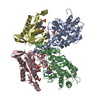
Mass: 96.063 Da / Num. of mol.: 4 / Source method: obtained synthetically / Formula: SO4
#4: Chemical ChemComp-AMP / ADENOSINE MONOPHOSPHATE


Mass: 347.221 Da / Num. of mol.: 2 / Source method: obtained synthetically / Formula: C10H14N5O7P / Comment: AMP*YM
#5: Chemical ChemComp-GDP / GUANOSINE-5'-DIPHOSPHATE


Type: RNA linking / Mass: 443.201 Da / Num. of mol.: 2 / Source method: obtained synthetically / Formula: C10H15N5O11P2 / Comment: GDP, energy-carrying molecule*YM
#6: Chemical ChemComp-MG / MAGNESIUM ION


Mass: 24.305 Da / Num. of mol.: 2 / Source method: obtained synthetically / Formula: Mg
#7: Water ChemComp-HOH / water


Mass: 18.015 Da / Num. of mol.: 829 / Source method: isolated from a natural source / Formula: H2O

-
Experimental details

-
Experiment

ExperimentMethod: X-RAY DIFFRACTION / Number of used crystals: 1

-
Sample preparation

CrystalDensity Matthews: 2.43 Å3/Da / Density % sol: 49.37 %
Crystal growTemperature: 277 K / Method: vapor diffusion, sitting drop / pH: 5.5
Details: PEG 3350, Bis-Tris, (NH4)2SO4, pH 5.5, VAPOR DIFFUSION, SITTING DROP, temperature 277.0K

-
Data collection

DiffractionMean temperature: 100 K
Diffraction sourceSource: SYNCHROTRON / Site: ALS / Beamline: 8.2.1 / Wavelength: 1 Å
DetectorType: ADSC QUANTUM 315r / Detector: CCD / Date: Dec 19, 2009
RadiationMonochromator: Double crystal, Si(111) / Protocol: SINGLE WAVELENGTH / Monochromatic (M) / Laue (L): M / Scattering type: x-ray
Radiation wavelengthWavelength: 1 Å / Relative weight: 1
ReflectionResolution: 2.3→50 Å / Num. obs: 49917 / % possible obs: 99.7 % / Redundancy: 6.8 % / Rmerge(I) obs: 0.117
Reflection shellResolution: 2.3→2.38 Å / Redundancy: 4 % / Rmerge(I) obs: 0.447 / Mean I/σ(I) obs: 2 / Num. unique all: 4766 / % possible all: 97.3

-
Processing

SoftwareName: PHENIX / Version: (phenix.refine: 1.8.1_1168) / Classification: refinement
RefinementMethod to determine structure: MOLECULAR REPLACEMENT / Resolution: 2.3→44.955 Å / SU ML: 0.18 / σ(F): 1.34 / Phase error: 21.18 / Stereochemistry target values: ML
RfactorNum. reflection% reflectionSelection details
Rfree0.2255 2524 5.07 %random
Rwork0.1781 ---
obs0.1806 49826 99.59 %-
Solvent computationShrinkage radii: 0.9 Å / VDW probe radii: 1.11 Å / Solvent model: FLAT BULK SOLVENT MODEL
Refinement stepCycle: LAST / Resolution: 2.3→44.955 Å
ProteinNucleic acidLigandSolventTotal
Num. atoms7431 0 124 829 8384
Refine LS restraints
Refine-IDTypeDev idealNumber
X-RAY DIFFRACTIONf_bond_d0.0047684
X-RAY DIFFRACTIONf_angle_d0.85610422
X-RAY DIFFRACTIONf_dihedral_angle_d15.3792922
X-RAY DIFFRACTIONf_chiral_restr0.0581212
X-RAY DIFFRACTIONf_plane_restr0.0041329
LS refinement shell
Resolution (Å)Rfactor RfreeNum. reflection RfreeRfactor RworkNum. reflection RworkRefine-ID% reflection obs (%)
2.3003-2.34450.27711370.22442444X-RAY DIFFRACTION94
2.3445-2.39240.27411430.20922564X-RAY DIFFRACTION99
2.3924-2.44440.26071440.19812597X-RAY DIFFRACTION100
2.4444-2.50130.25431230.19522573X-RAY DIFFRACTION100
2.5013-2.56380.27421510.19982624X-RAY DIFFRACTION100
2.5638-2.63310.24491310.19572620X-RAY DIFFRACTION100
2.6331-2.71060.25241430.19082601X-RAY DIFFRACTION100
2.7106-2.79810.23791350.19362613X-RAY DIFFRACTION100
2.7981-2.89810.29131490.19232616X-RAY DIFFRACTION100
2.8981-3.01410.24071410.19382625X-RAY DIFFRACTION100
3.0141-3.15120.24661460.19252619X-RAY DIFFRACTION100
3.1512-3.31730.2631260.17842622X-RAY DIFFRACTION100
3.3173-3.52510.22281420.1752661X-RAY DIFFRACTION100
3.5251-3.79710.21981350.16672648X-RAY DIFFRACTION100
3.7971-4.1790.17761380.14962660X-RAY DIFFRACTION100
4.179-4.78310.16841300.14122699X-RAY DIFFRACTION100
4.7831-6.02390.20251460.17492701X-RAY DIFFRACTION100
6.0239-44.96320.18891640.17542815X-RAY DIFFRACTION99

+
About Yorodumi

-
News

-
Feb 9, 2022. New format data for meta-information of EMDB entries

New format data for meta-information of EMDB entries

  • Version 3 of the EMDB header file is now the official format.
  • The previous official version 1.9 will be removed from the archive.

Related info.:EMDB header

External links:wwPDB to switch to version 3 of the EMDB data model

-
Aug 12, 2020. Covid-19 info

Covid-19 info

URL: https://pdbjlvh1.pdbj.org/emnavi/covid19.php

New page: Covid-19 featured information page in EM Navigator.

Related info.:Covid-19 info / Mar 5, 2020. Novel coronavirus structure data

+
Mar 5, 2020. Novel coronavirus structure data

Novel coronavirus structure data

Related info.:Yorodumi Speices / Aug 12, 2020. Covid-19 info

External links:COVID-19 featured content - PDBj / Molecule of the Month (242):Coronavirus Proteases

+
Jan 31, 2019. EMDB accession codes are about to change! (news from PDBe EMDB page)

EMDB accession codes are about to change! (news from PDBe EMDB page)

  • The allocation of 4 digits for EMDB accession codes will soon come to an end. Whilst these codes will remain in use, new EMDB accession codes will include an additional digit and will expand incrementally as the available range of codes is exhausted. The current 4-digit format prefixed with “EMD-” (i.e. EMD-XXXX) will advance to a 5-digit format (i.e. EMD-XXXXX), and so on. It is currently estimated that the 4-digit codes will be depleted around Spring 2019, at which point the 5-digit format will come into force.
  • The EM Navigator/Yorodumi systems omit the EMD- prefix.

Related info.:Q: What is EMD? / ID/Accession-code notation in Yorodumi/EM Navigator

External links:EMDB Accession Codes are Changing Soon! / Contact to PDBj

+
Jul 12, 2017. Major update of PDB

Major update of PDB

  • wwPDB released updated PDB data conforming to the new PDBx/mmCIF dictionary.
  • This is a major update changing the version number from 4 to 5, and with Remediation, in which all the entries are updated.
  • In this update, many items about electron microscopy experimental information are reorganized (e.g. em_software).
  • Now, EM Navigator and Yorodumi are based on the updated data.

External links:wwPDB Remediation / Enriched Model Files Conforming to OneDep Data Standards Now Available in the PDB FTP Archive

-
Yorodumi

Thousand views of thousand structures

  • Yorodumi is a browser for structure data from EMDB, PDB, SASBDB, etc.
  • This page is also the successor to EM Navigator detail page, and also detail information page/front-end page for Omokage search.
  • The word "yorodu" (or yorozu) is an old Japanese word meaning "ten thousand". "mi" (miru) is to see.

Related info.:EMDB / PDB / SASBDB / Comparison of 3 databanks / Yorodumi Search / Aug 31, 2016. New EM Navigator & Yorodumi / Yorodumi Papers / Jmol/JSmol / Function and homology information / Changes in new EM Navigator and Yorodumi

Read more