Mass: 18.015 Da / Num. of mol.: 204 / Source method: isolated from a natural source / Formula: H2O
-
Experimental details
-
Experiment
Experiment
Method: X-RAY DIFFRACTION / Number of used crystals: 1
-
Sample preparation
Crystal
Density Matthews: 2.67 Å3/Da / Density % sol: 53.89 %
Crystal grow
Temperature: 277 K / Method: vapor diffusion, sitting drop / pH: 6.5 Details: PEG 550 MME, MES, ZnSO4, pH 6.5, temperature 277K, VAPOR DIFFUSION, SITTING DROP
-
Data collection
Diffraction
ID
Mean temperature (K)
Crystal-ID
1
100
1
2
100
1
Diffraction source
Source
Site
Beamline
ID
Wavelength (Å)
SYNCHROTRON
ALS
8.2.1
1
1
SYNCHROTRON
ALS
8.2.1
2
1.2824
Detector
Type
ID
Detector
Date
ADSC QUANTUM 315r
1
CCD
Jun 30, 2009
ADSC QUANTUM 315r
2
CCD
Jun 30, 2009
Radiation
ID
Protocol
Monochromatic (M) / Laue (L)
Scattering type
Wavelength-ID
1
SINGLEWAVELENGTH
M
x-ray
1
2
SINGLEWAVELENGTH
M
x-ray
1
Radiation wavelength
ID
Wavelength (Å)
Relative weight
1
1
1
2
1.2824
1
Reflection
Redundancy: 10.3 % / Av σ(I) over netI: 52.04 / Number: 257894 / Rmerge(I) obs: 0.061 / Χ2: 2.12 / D res high: 2 Å / D res low: 50 Å / Num. obs: 24925 / % possible obs: 99.9
Diffraction reflection shell
Highest resolution (Å)
Lowest resolution (Å)
% possible obs (%)
ID
Rmerge(I) obs
Chi squared
Redundancy
5.43
50
98.4
1
0.05
5.543
9.4
4.31
5.43
99.9
1
0.049
4.179
9.9
3.76
4.31
100
1
0.051
3.631
9.7
3.42
3.76
100
1
0.058
3.746
9.7
3.17
3.42
100
1
0.063
3.48
10
2.99
3.17
100
1
0.066
2.989
10.2
2.84
2.99
100
1
0.069
2.608
10.3
2.71
2.84
100
1
0.078
2.242
10.5
2.61
2.71
100
1
0.081
1.828
10.5
2.52
2.61
100
1
0.093
1.69
10.6
2.44
2.52
100
1
0.102
1.482
10.6
2.37
2.44
100
1
0.112
1.311
10.6
2.31
2.37
100
1
0.12
1.209
10.7
2.25
2.31
100
1
0.136
1.138
10.6
2.2
2.25
100
1
0.155
1.098
10.6
2.15
2.2
100
1
0.18
1.017
10.7
2.11
2.15
100
1
0.236
0.964
10.6
2.07
2.11
100
1
0.28
0.898
10.7
2.03
2.07
100
1
0.3
0.913
10.6
2
2.03
100
1
0.355
0.833
10.6
Reflection
Resolution: 1.85→50 Å / Num. obs: 31631 / % possible obs: 99.7 % / Redundancy: 11.2 % / Rmerge(I) obs: 0.044 / Χ2: 1.026 / Net I/σ(I): 14.7
Reflection shell
Resolution: 1.85→1.88 Å / Redundancy: 9 % / Rmerge(I) obs: 0.606 / Mean I/σ(I) obs: 3 / Num. unique all: 1527 / Χ2: 0.654 / % possible all: 98.5
-
Phasing
Phasing
Method: SAD
-
Processing
Software
Name
Version
Classification
NB
DENZO
datareduction
SCALEPACK
datascaling
PHENIX
1.5_2
refinement
PDB_EXTRACT
3.1
dataextraction
AutoSol
phasing
Refinement
Method to determine structure: SAD / Resolution: 1.846→31.471 Å / Occupancy max: 1 / Occupancy min: 1 / FOM work R set: 0.814 / SU ML: 0.26 / σ(F): 1.33 / Phase error: 24.89 / Stereochemistry target values: ML
Rfactor
Num. reflection
% reflection
Rfree
0.242
1592
5.04 %
Rwork
0.212
-
-
obs
0.213
31559
99.56 %
Solvent computation
Shrinkage radii: 0.9 Å / VDW probe radii: 1.11 Å / Solvent model: FLAT BULK SOLVENT MODEL / Bsol: 53.234 Å2 / ksol: 0.335 e/Å3
In the structure databanks used in Yorodumi, some data are registered as the other names, "COVID-19 virus" and "2019-nCoV". Here are the details of the virus and the list of structure data.
Jan 31, 2019. EMDB accession codes are about to change! (news from PDBe EMDB page)
EMDB accession codes are about to change! (news from PDBe EMDB page)
The allocation of 4 digits for EMDB accession codes will soon come to an end. Whilst these codes will remain in use, new EMDB accession codes will include an additional digit and will expand incrementally as the available range of codes is exhausted. The current 4-digit format prefixed with “EMD-” (i.e. EMD-XXXX) will advance to a 5-digit format (i.e. EMD-XXXXX), and so on. It is currently estimated that the 4-digit codes will be depleted around Spring 2019, at which point the 5-digit format will come into force.
The EM Navigator/Yorodumi systems omit the EMD- prefix.
Related info.:Q: What is EMD? / ID/Accession-code notation in Yorodumi/EM Navigator
Yorodumi is a browser for structure data from EMDB, PDB, SASBDB, etc.
This page is also the successor to EM Navigator detail page, and also detail information page/front-end page for Omokage search.
The word "yorodu" (or yorozu) is an old Japanese word meaning "ten thousand". "mi" (miru) is to see.
Related info.:EMDB / PDB / SASBDB / Comparison of 3 databanks / Yorodumi Search / Aug 31, 2016. New EM Navigator & Yorodumi / Yorodumi Papers / Jmol/JSmol / Function and homology information / Changes in new EM Navigator and Yorodumi
 Movie
Movie Controller


 Open data
 Basic information
 Components
 Keywords
 Function and homology information
 Histophilus somni (bacteria)
 X-RAY DIFFRACTION /
  Authors
 Citation
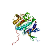
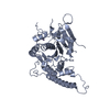
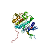
 Structure visualization
 Downloads & links
 Other downloads
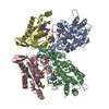


 PDBj
PDBj Assembly

 Mass: 65.409 Da / Num. of mol.: 2 / Source method: obtained synthetically / Formula: Zn
 Mass: 96.063 Da / Num. of mol.: 1 / Source method: obtained synthetically / Formula: SO4
 Mass: 18.015 Da / Num. of mol.: 204 / Source method: isolated from a natural source / Formula: H2O
 Sample preparation
 Processing