 Movie
Movie Controller


+ Open data

- Basic information
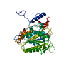
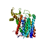
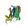
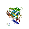
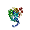
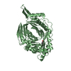
| Entry | Database: PDB / ID: 1toc | ||||||
|---|---|---|---|---|---|---|---|
| Title | STRUCTURE OF SERINE PROTEINASE | ||||||
 Components Components |
| ||||||
 Keywords Keywords | COMPLEX (HYDROLASE/INHIBITOR) / VITAMIN K / ZYMOGEN / GAMMA-CARBOXYGLUTAMIC ACID / ACUTE PHASE / LIVER / HYDROLASE / SERINE PROTEASE KUNITZ-LIKE INHIBITOR / KRINGLE / COMPLEX (HYDROLASE-INHIBITOR) COMPLEX | ||||||
| Function / homology |  Function and homology information Function and homology informationfibrinogen binding / thrombin / positive regulation of blood coagulation / protein polymerization / transport vesicle / acute-phase response / serine-type endopeptidase inhibitor activity / platelet activation / toxin activity / serine-type endopeptidase activity ...fibrinogen binding / thrombin / positive regulation of blood coagulation / protein polymerization / transport vesicle / acute-phase response / serine-type endopeptidase inhibitor activity / platelet activation / toxin activity / serine-type endopeptidase activity / calcium ion binding / proteolysis / : / extracellular region Similarity search - Function | ||||||
| Biological species |   Ornithodoros moubata (arthropod) Ornithodoros moubata (arthropod) | ||||||
| Method |  X-RAY DIFFRACTION / Resolution: 3.1 Å X-RAY DIFFRACTION / Resolution: 3.1 Å | ||||||
 Authors Authors | Van De Locht, A. / Huber, R. / Bode, W. | ||||||
 Citation Citation | Journal: EMBO J. / Year: 1996 Title: The ornithodorin-thrombin crystal structure, a key to the TAP enigma? Authors: van de Locht, A. / Stubbs, M.T. / Bode, W. / Friedrich, T. / Bollschweiler, C. / Hoffken, W. / Huber, R. #1: Journal: Embo J. / Year: 1995Title: Two Heads are Better Than One: Crystal Structure of the Insect Derived Double Domain Kazal Inhibitor Rhodniin in Complex with Thrombin Authors: Van De Locht, A. / Lamba, D. / Bauer, M. / Huber, R. / Friedrich, T. / Kroger, B. / Hoffken, W. / Bode, W. #2: Journal: Embo J. / Year: 1989Title: The Refined 1.9 A Crystal Structure of Human Alpha-Thrombin: Interaction with D-Phe-Pro-Arg Chloromethylketone and Significance of the Tyr-Pro-Pro-Trp Insertion Segment Authors: Bode, W. / Mayr, I. / Baumann, U. / Huber, R. / Stone, S.R. / Hofsteenge, J. | ||||||
| History |
|
- Structure visualization
| Structure viewer | Molecule: MolmilJmol/JSmol |
|---|
- Downloads & links
-Download
| PDBx/mmCIF format | 1toc.cif.gz | 334.1 KB | Display | PDBx/mmCIF format |
|---|---|---|---|---|
| PDB format | pdb1toc.ent.gz | 271.5 KB | Display | PDB format |
| PDBx/mmJSON format | 1toc.json.gz | Tree view | PDBx/mmJSON format | |
| Others |  Other downloads Other downloads |
-Validation report
| Arichive directory | https://data.pdbj.org/pub/pdb/validation_reports/to/1tocftp://data.pdbj.org/pub/pdb/validation_reports/to/1toc | HTTPS FTP |
|---|
-Related structure data
| Similar structure data |
|---|


-Links
 PDBj
PDBj


- Assembly
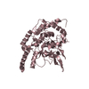
| Deposited unit | 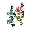
| ||||||||
|---|---|---|---|---|---|---|---|---|---|
| 1 | 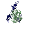
| ||||||||
| 2 | 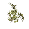
| ||||||||
| 3 | 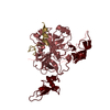
| ||||||||
| 4 | 
| ||||||||
| Unit cell |
|
-Components
| #1: Protein/peptide | Mass: 5735.240 Da / Num. of mol.: 4 Source method: isolated from a genetically manipulated source Source: (gene. exp.)  #2: Protein | Mass: 29772.422 Da / Num. of mol.: 4 Source method: isolated from a genetically manipulated source Source: (gene. exp.) #3: Protein | Mass: 12726.731 Da / Num. of mol.: 4 Source method: isolated from a genetically manipulated source Source: (gene. exp.) Ornithodoros moubata (arthropod) / Plasmid: PMD8A / Production host: #4: Water | ChemComp-HOH / |  Mass: 18.015 Da / Num. of mol.: 31 / Source method: isolated from a natural source / Formula: H2O Mass: 18.015 Da / Num. of mol.: 31 / Source method: isolated from a natural source / Formula: H2OHas protein modification | Y | |
|---|
-Experimental details
-Experiment
| Experiment | Method: X-RAY DIFFRACTION |
|---|
- Sample preparation
| Crystal | Density Matthews: 2.81 Å3/Da / Density % sol: 56 % | |||||||||||||||||||||||||
|---|---|---|---|---|---|---|---|---|---|---|---|---|---|---|---|---|---|---|---|---|---|---|---|---|---|---|
| Crystal grow | *PLUS Temperature: 20 ℃ / pH: 5 / Method: vapor diffusion, hanging drop | |||||||||||||||||||||||||
| Components of the solutions | *PLUS
|
-Data collection
| Diffraction source | Wavelength: 1.5418 |
|---|---|
| Detector | Type: MARRESEARCH / Detector: IMAGE PLATE / Date: Jan 13, 1996 |
| Radiation | Monochromatic (M) / Laue (L): M / Scattering type: x-ray |
| Radiation wavelength | Wavelength: 1.5418 Å / Relative weight: 1 |
| Reflection | Num. obs: 94484 / % possible obs: 89 % / Observed criterion σ(I): 3 / Redundancy: 2.39 % / Rmerge(I) obs: 0.14 |
| Reflection | *PLUS Highest resolution: 3 Å / Lowest resolution: 9999 Å |
| Reflection shell | *PLUS Highest resolution: 3 Å / Lowest resolution: 3.13 Å / % possible obs: 75.2 % |
- Processing
| Software |
| ||||||||||||||||||||||||||||||||||||||||||||||||||||||||||||
|---|---|---|---|---|---|---|---|---|---|---|---|---|---|---|---|---|---|---|---|---|---|---|---|---|---|---|---|---|---|---|---|---|---|---|---|---|---|---|---|---|---|---|---|---|---|---|---|---|---|---|---|---|---|---|---|---|---|---|---|---|---|
| Refinement | Resolution: 3.1→10 Å / σ(F): 2 /
| ||||||||||||||||||||||||||||||||||||||||||||||||||||||||||||
| Refinement step | Cycle: LAST / Resolution: 3.1→10 Å
| ||||||||||||||||||||||||||||||||||||||||||||||||||||||||||||
| Refine LS restraints |
| ||||||||||||||||||||||||||||||||||||||||||||||||||||||||||||
| Software | *PLUS Name: X-PLOR / Classification: refinement | ||||||||||||||||||||||||||||||||||||||||||||||||||||||||||||
| Refinement | *PLUS | ||||||||||||||||||||||||||||||||||||||||||||||||||||||||||||
| Solvent computation | *PLUS | ||||||||||||||||||||||||||||||||||||||||||||||||||||||||||||
| Displacement parameters | *PLUS |
