 Movie
Movie Controller

[English] 日本語
 Yorodumi
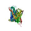
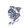
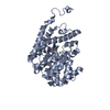
- PDB-1iyl: Crystal Structure of Candida albicans N-myristoyltransferase with... -
+ Open data

- Basic information
| Entry | Database: PDB / ID: 1iyl | ||||||
|---|---|---|---|---|---|---|---|
| Title | Crystal Structure of Candida albicans N-myristoyltransferase with Non-peptidic Inhibitor | ||||||
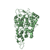
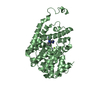
 Components Components | Myristoyl-CoA:Protein N-Myristoyltransferase | ||||||
 Keywords Keywords | TRANSFERASE | ||||||
| Function / homology |  Function and homology information Function and homology informationglycylpeptide N-tetradecanoyltransferase / glycylpeptide N-tetradecanoyltransferase activity / protein localization to membrane / cytosol Similarity search - Function | ||||||
| Biological species |  Candida albicans (yeast) Candida albicans (yeast) | ||||||
| Method |  X-RAY DIFFRACTION / SYNCHROTRON / MOLECULAR REPLACEMENT / Resolution: 3.2 Å X-RAY DIFFRACTION / SYNCHROTRON / MOLECULAR REPLACEMENT / Resolution: 3.2 Å | ||||||
 Authors Authors | Sogabe, S. / Fukami, T.A. / Morikami, K. / Shiratori, Y. / Aoki, Y. / D'Arcy, A. / Winkler, F.K. / Banner, D.W. / Ohtsuka, T. | ||||||
 Citation Citation | Journal: CHEM.BIOL. / Year: 2002 Title: Crystal Structures of Candida albicans N-Myristoyltransferase with Two Distinct Inhibitors Authors: Sogabe, S. / Masubuchi, M. / Sakata, K. / Fukami, T.A. / Morikami, K. / Shiratori, Y. / Ebiike, H. / Kawasaki, K. / Aoki, Y. / Shimma, N. / D'Arcy, A. / Winkler, F.K. / Banner, D.W. / Ohtsuka, T. | ||||||
| History |
|
- Structure visualization
| Structure viewer | Molecule: MolmilJmol/JSmol |
|---|
- Downloads & links
-Download
| PDBx/mmCIF format | 1iyl.cif.gz | 311.9 KB | Display | PDBx/mmCIF format |
|---|---|---|---|---|
| PDB format | pdb1iyl.ent.gz | 253.2 KB | Display | PDB format |
| PDBx/mmJSON format | 1iyl.json.gz | Tree view | PDBx/mmJSON format | |
| Others |  Other downloads Other downloads |
-Validation report
| Arichive directory | https://data.pdbj.org/pub/pdb/validation_reports/iy/1iylftp://data.pdbj.org/pub/pdb/validation_reports/iy/1iyl | HTTPS FTP |
|---|
-Related structure data
| Related structure data | 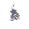 1iykSC S: Starting model for refinement C: citing same article ( |
|---|---|
| Similar structure data |


-Links
 PDBj
PDBj

- Assembly
| Deposited unit | 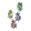
| ||||||||
|---|---|---|---|---|---|---|---|---|---|
| 1 | 
| ||||||||
| 2 | 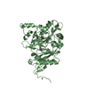
| ||||||||
| 3 | 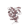
| ||||||||
| 4 | 
| ||||||||
| 5 | 
| ||||||||
| 6 | 
| ||||||||
| Unit cell |
|
-Components
| #1: Protein | Mass: 45421.566 Da / Num. of mol.: 4 Source method: isolated from a genetically manipulated source Source: (gene. exp.) Candida albicans (yeast) / Plasmid: pKF19 / Production host:  References: UniProt: P30418, glycylpeptide N-tetradecanoyltransferase #2: Chemical | 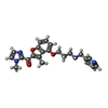  Mass: 404.462 Da / Num. of mol.: 3 / Source method: obtained synthetically / Formula: C23H24N4O3 Mass: 404.462 Da / Num. of mol.: 3 / Source method: obtained synthetically / Formula: C23H24N4O3 |
|---|
-Experimental details
-Experiment
| Experiment | Method: X-RAY DIFFRACTION / Number of used crystals: 1 |
|---|
- Sample preparation
| Crystal | Density Matthews: 3.35 Å3/Da / Density % sol: 63.25 % | ||||||||||||||||||||||||||||||
|---|---|---|---|---|---|---|---|---|---|---|---|---|---|---|---|---|---|---|---|---|---|---|---|---|---|---|---|---|---|---|---|
| Crystal grow | Temperature: 277 K / Method: vapor diffusion, hanging drop / pH: 7.5 Details: PEG3350, lithium sulfate, HEPES, pH 7.5, VAPOR DIFFUSION, HANGING DROP, temperature 277K | ||||||||||||||||||||||||||||||
| Crystal grow | *PLUS Temperature: 4 ℃ | ||||||||||||||||||||||||||||||
| Components of the solutions | *PLUS
|
-Data collection
| Diffraction | Mean temperature: 288 K |
|---|---|
| Diffraction source | Source: SYNCHROTRON / Site: Photon Factory  / Beamline: BL-6B / Wavelength: 1 Å / Beamline: BL-6B / Wavelength: 1 Å |
| Detector | Type: WEISSENBERG / Detector: DIFFRACTOMETER / Date: Nov 28, 1998 |
| Radiation | Monochromator: Si(111) / Protocol: SINGLE WAVELENGTH / Monochromatic (M) / Laue (L): M / Scattering type: x-ray |
| Radiation wavelength | Wavelength: 1 Å / Relative weight: 1 |
| Reflection | Resolution: 3.2→50 Å / Num. all: 40711 / Num. obs: 40711 / % possible obs: 99 % / Observed criterion σ(F): 0 / Observed criterion σ(I): 0 / Redundancy: 3.5 % / Rmerge(I) obs: 0.072 / Net I/σ(I): 16.4 |
| Reflection shell | Resolution: 3.2→3.31 Å / Redundancy: 3.5 % / Rmerge(I) obs: 0.364 / Mean I/σ(I) obs: 3.1 / Num. unique all: 4020 / % possible all: 99.9 |
| Reflection | *PLUS Lowest resolution: 50 Å |
- Processing
| Software |
| ||||||||||||||||||||||||||||||||||||
|---|---|---|---|---|---|---|---|---|---|---|---|---|---|---|---|---|---|---|---|---|---|---|---|---|---|---|---|---|---|---|---|---|---|---|---|---|---|
| Refinement | Method to determine structure: MOLECULAR REPLACEMENT Starting model: PDB ENTRY 1IYK Resolution: 3.2→40 Å / Rfactor Rfree error: 0.008 / Isotropic thermal model: B-GROUP / Cross valid method: THROUGHOUT / σ(F): 0 / Stereochemistry target values: Engh & Huber
| ||||||||||||||||||||||||||||||||||||
| Solvent computation | Solvent model: FLAT MODEL / Bsol: 53.4476 Å2 / ksol: 0.232096 e/Å3 | ||||||||||||||||||||||||||||||||||||
| Displacement parameters | Biso mean: 45.4 Å2 | ||||||||||||||||||||||||||||||||||||
| Refine analyze |
| ||||||||||||||||||||||||||||||||||||
| Refinement step | Cycle: LAST / Resolution: 3.2→40 Å
| ||||||||||||||||||||||||||||||||||||
| Refine LS restraints |
| ||||||||||||||||||||||||||||||||||||
| LS refinement shell | Resolution: 3.2→3.31 Å / Rfactor Rfree error: 0.024 / Total num. of bins used: 10
| ||||||||||||||||||||||||||||||||||||
| Refinement | *PLUS Lowest resolution: 40 Å / % reflection Rfree: 5 % | ||||||||||||||||||||||||||||||||||||
| Solvent computation | *PLUS | ||||||||||||||||||||||||||||||||||||
| Displacement parameters | *PLUS | ||||||||||||||||||||||||||||||||||||
| Refine LS restraints | *PLUS
| ||||||||||||||||||||||||||||||||||||
| LS refinement shell | *PLUS Highest resolution: 3.2 Å |
