 Movie
Movie Controller

[English] 日本語
 Yorodumi
- PDB-1no7: Structure of the Large Protease Resistant Upper Domain of VP5, th... -
+ Open data

- Basic information
| Entry | Database: PDB / ID: 1no7 | ||||||
|---|---|---|---|---|---|---|---|
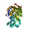
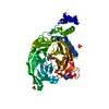
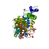
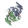
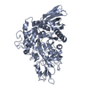
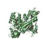
| Title | Structure of the Large Protease Resistant Upper Domain of VP5, the Major Capsid Protein of Herpes Simplex Virus-1 | ||||||
 Components Components | Major capsid protein | ||||||
 Keywords Keywords | VIRAL PROTEIN / novel fold / alpha plus beta / viral capsid protein / folding nucleus | ||||||
| Function / homology | T=16 icosahedral viral capsid / Herpesvirus major capsid protein / Herpesvirus major capsid protein, upper domain superfamily / Herpes virus major capsid protein / host cell nucleus / structural molecule activity / Major capsid protein Function and homology information Function and homology information | ||||||
| Biological species |   Human herpesvirus 1 (Herpes simplex virus type 1) Human herpesvirus 1 (Herpes simplex virus type 1) | ||||||
| Method |  X-RAY DIFFRACTION / SYNCHROTRON / MAD / Resolution: 2.9 Å X-RAY DIFFRACTION / SYNCHROTRON / MAD / Resolution: 2.9 Å | ||||||
 Authors Authors | Bowman, B.R. / Baker, M.L. / Rixon, F.J. / Chiu, W. / Quiocho, F.A. | ||||||
 Citation Citation | Journal: Embo J. / Year: 2003 Title: Structure of the herpesvirus major capsid protein Authors: Bowman, B.R. / Baker, M.L. / Rixon, F.J. / Chiu, W. / Quiocho, F.A. | ||||||
| History |
|
- Structure visualization
| Structure viewer | Molecule: MolmilJmol/JSmol |
|---|
- Downloads & links
-Download
| PDBx/mmCIF format | 1no7.cif.gz | 195.9 KB | Display | PDBx/mmCIF format |
|---|---|---|---|---|
| PDB format | pdb1no7.ent.gz | 157.1 KB | Display | PDB format |
| PDBx/mmJSON format | 1no7.json.gz | Tree view | PDBx/mmJSON format | |
| Others |  Other downloads Other downloads |
-Validation report
| Arichive directory | https://data.pdbj.org/pub/pdb/validation_reports/no/1no7ftp://data.pdbj.org/pub/pdb/validation_reports/no/1no7 | HTTPS FTP |
|---|
-Related structure data
| Similar structure data |
|---|


-Links
 PDBj
PDBj
- Assembly
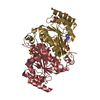
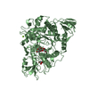
| Deposited unit | 
| ||||||||
|---|---|---|---|---|---|---|---|---|---|
| 1 | 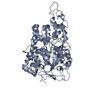
| ||||||||
| 2 | 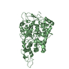
| ||||||||
| Unit cell |
|
-Components
| #1: Protein | Mass: 65169.766 Da / Num. of mol.: 2 / Fragment: VP5 upper domain Source method: isolated from a genetically manipulated source Source: (gene. exp.) Human herpesvirus 1 (Herpes simplex virus type 1)Genus: Simplexvirus / Gene: UL19 / Plasmid: pET32 / Species (production host): Escherichia coli / Production host:  |
|---|
-Experimental details
-Experiment
| Experiment | Method: X-RAY DIFFRACTION / Number of used crystals: 2 |
|---|
- Sample preparation
| Crystal | Density Matthews: 4.87 Å3/Da / Density % sol: 74.55 % | |||||||||||||||||||||||||||||||||||||||||||||||||
|---|---|---|---|---|---|---|---|---|---|---|---|---|---|---|---|---|---|---|---|---|---|---|---|---|---|---|---|---|---|---|---|---|---|---|---|---|---|---|---|---|---|---|---|---|---|---|---|---|---|---|
| Crystal grow | Temperature: 277 K / Method: vapor diffusion, hanging drop / pH: 6.2 Details: sodium acetate, imidazole, pH 6.2, VAPOR DIFFUSION, HANGING DROP, temperature 277K | |||||||||||||||||||||||||||||||||||||||||||||||||
| Crystal grow | *PLUS Temperature: 4 ℃ / pH: 8 / Method: vapor diffusion | |||||||||||||||||||||||||||||||||||||||||||||||||
| Components of the solutions | *PLUS
|
-Data collection
| Diffraction |
| |||||||||||||||
|---|---|---|---|---|---|---|---|---|---|---|---|---|---|---|---|---|
| Diffraction source |
| |||||||||||||||
| Detector |
| |||||||||||||||
| Radiation |
| |||||||||||||||
| Radiation wavelength |
| |||||||||||||||
| Reflection | Resolution: 2.9→50 Å / Num. obs: 47194 / % possible obs: 91.2 % / Biso Wilson estimate: 31 Å2 / Net I/σ(I): 7 | |||||||||||||||
| Reflection shell | Highest resolution: 2.9 Å / Mean I/σ(I) obs: 3.3 / % possible all: 80.4 | |||||||||||||||
| Reflection | *PLUS Num. measured all: 308549 / Rmerge(I) obs: 0.101 | |||||||||||||||
| Reflection shell | *PLUS % possible obs: 80.4 % / Rmerge(I) obs: 0.205 |

- Processing
| Software |
| ||||||||||||||||||||||||||||||||||||
|---|---|---|---|---|---|---|---|---|---|---|---|---|---|---|---|---|---|---|---|---|---|---|---|---|---|---|---|---|---|---|---|---|---|---|---|---|---|
| Refinement | Method to determine structure: MAD / Resolution: 2.9→47.09 Å / Rfactor Rfree error: 0.004 / Data cutoff high absF: 408294 / Data cutoff low absF: 0 / Isotropic thermal model: RESTRAINED / Cross valid method: THROUGHOUT / σ(F): 0
| ||||||||||||||||||||||||||||||||||||
| Solvent computation | Solvent model: FLAT MODEL / Bsol: 37.194 Å2 / ksol: 0.291374 e/Å3 | ||||||||||||||||||||||||||||||||||||
| Displacement parameters | Biso mean: 62.2 Å2
| ||||||||||||||||||||||||||||||||||||
| Refine analyze |
| ||||||||||||||||||||||||||||||||||||
| Refinement step | Cycle: LAST / Resolution: 2.9→47.09 Å
| ||||||||||||||||||||||||||||||||||||
| Refine LS restraints |
| ||||||||||||||||||||||||||||||||||||
| LS refinement shell | Resolution: 2.9→3.08 Å / Rfactor Rfree error: 0.016 / Total num. of bins used: 6
| ||||||||||||||||||||||||||||||||||||
| Xplor file |
| ||||||||||||||||||||||||||||||||||||
| Refinement | *PLUS Highest resolution: 2.9 Å / Lowest resolution: 50 Å / % reflection Rfree: 10 % / Rfactor Rwork: 0.254 | ||||||||||||||||||||||||||||||||||||
| Solvent computation | *PLUS | ||||||||||||||||||||||||||||||||||||
| Displacement parameters | *PLUS | ||||||||||||||||||||||||||||||||||||
| Refine LS restraints | *PLUS
|
