 Movie
Movie Controller

[English] 日本語
 Yorodumi
- PDB-6s9u: Crystal structure of sucrose 6F-phosphate phosphorylase from Ilum... -
+ Open data

- Basic information
| Entry | Database: PDB / ID: 6s9u | ||||||
|---|---|---|---|---|---|---|---|
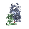
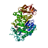
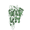
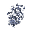
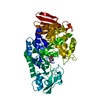
| Title | Crystal structure of sucrose 6F-phosphate phosphorylase from Ilumatobacter coccineus | ||||||
 Components Components | Putative sucrose phosphorylase | ||||||
 Keywords Keywords | TRANSFERASE / Glycoside phosphorylase / sucrose 6-phosphate / glycoside hydrolase family GH13-18 | ||||||
| Function / homology | sucrose 6F-phosphate phosphorylase / Oligo-1,6-glucosidase, domain 2 / glycosyltransferase activity / Glycoside hydrolase superfamily / carbohydrate metabolic process / PHOSPHATE ION / Sucrose 6(F)-phosphate phosphorylase / Sucrose 6(F)-phosphate phosphorylase Function and homology information Function and homology information | ||||||
| Biological species |  Ilumatobacter coccineus YM16-304 (bacteria) Ilumatobacter coccineus YM16-304 (bacteria) | ||||||
| Method |  X-RAY DIFFRACTION / SYNCHROTRON / MOLECULAR REPLACEMENT / molecular replacement / Resolution: 2.05 Å X-RAY DIFFRACTION / SYNCHROTRON / MOLECULAR REPLACEMENT / molecular replacement / Resolution: 2.05 Å | ||||||
 Authors Authors | Capra, N. / Franceus, J. / Desmet, T. / Thunnissen, A.M.W.H. | ||||||
 Citation Citation | Journal: Int J Mol Sci / Year: 2019 Title: Structural Comparison of a Promiscuous and a Highly Specific Sucrose 6 F -Phosphate Phosphorylase. Authors: Franceus, J. / Capra, N. / Desmet, T. / Thunnissen, A.W.H. | ||||||
| History |
|
- Structure visualization
| Structure viewer | Molecule: MolmilJmol/JSmol |
|---|
- Downloads & links
-Download
| PDBx/mmCIF format | 6s9u.cif.gz | 308.5 KB | Display | PDBx/mmCIF format |
|---|---|---|---|---|
| PDB format | pdb6s9u.ent.gz | 256.2 KB | Display | PDB format |
| PDBx/mmJSON format | 6s9u.json.gz | Tree view | PDBx/mmJSON format | |
| Others |  Other downloads Other downloads |
-Validation report
| Arichive directory | https://data.pdbj.org/pub/pdb/validation_reports/s9/6s9uftp://data.pdbj.org/pub/pdb/validation_reports/s9/6s9u | HTTPS FTP |
|---|
-Related structure data


-Links
 PDBj
PDBj- Assembly
| Deposited unit | 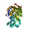
| ||||||||
|---|---|---|---|---|---|---|---|---|---|
| 1 |
| ||||||||
| Unit cell |
|
-Components
| #1: Protein | Mass: 59966.914 Da / Num. of mol.: 1 Source method: isolated from a genetically manipulated source Source: (gene. exp.) Ilumatobacter coccineus YM16-304 (bacteria)Gene: YM304_32550 / Production host: References: UniProt: M5A566, UniProt: A0A6C7EEG6*PLUS, sucrose phosphorylase | ||||||
|---|---|---|---|---|---|---|---|
| #2: Chemical | ChemComp-PO4 /   Mass: 94.971 Da / Num. of mol.: 1 / Source method: obtained synthetically / Formula: PO4 Mass: 94.971 Da / Num. of mol.: 1 / Source method: obtained synthetically / Formula: PO4 | ||||||
| #3: Chemical |   Mass: 282.334 Da / Num. of mol.: 2 / Source method: obtained synthetically / Formula: C11H26N2O6 / Comment: pH buffer*YM Mass: 282.334 Da / Num. of mol.: 2 / Source method: obtained synthetically / Formula: C11H26N2O6 / Comment: pH buffer*YM#4: Chemical | ChemComp-TRS / |   Mass: 122.143 Da / Num. of mol.: 1 / Source method: obtained synthetically / Formula: C4H12NO3 / Comment: pH buffer*YM Mass: 122.143 Da / Num. of mol.: 1 / Source method: obtained synthetically / Formula: C4H12NO3 / Comment: pH buffer*YM#5: Water | ChemComp-HOH / |  Mass: 18.015 Da / Num. of mol.: 313 / Source method: isolated from a natural source / Formula: H2O Mass: 18.015 Da / Num. of mol.: 313 / Source method: isolated from a natural source / Formula: H2OHas ligand of interest | N | |
-Experimental details
-Experiment
| Experiment | Method: X-RAY DIFFRACTION / Number of used crystals: 1 |
|---|
- Sample preparation
| Crystal | Density Matthews: 2.19 Å3/Da / Density % sol: 43.85 % |
|---|---|
| Crystal grow | Temperature: 291 K / Method: vapor diffusion, sitting drop / pH: 7.5 Details: 20% PEG 3350, 200 mM Na/K phosphate, 100 mM Bis-Tris propane |
-Data collection
| Diffraction | Mean temperature: 100 K / Serial crystal experiment: N | ||||||||||||||||||||||||||||||
|---|---|---|---|---|---|---|---|---|---|---|---|---|---|---|---|---|---|---|---|---|---|---|---|---|---|---|---|---|---|---|---|
| Diffraction source | Source: SYNCHROTRON / Site: ESRF  / Beamline: MASSIF-3 / Wavelength: 0.9677 Å / Beamline: MASSIF-3 / Wavelength: 0.9677 Å | ||||||||||||||||||||||||||||||
| Detector | Type: DECTRIS EIGER X 4M / Detector: PIXEL / Date: Apr 19, 2018 | ||||||||||||||||||||||||||||||
| Radiation | Protocol: SINGLE WAVELENGTH / Monochromatic (M) / Laue (L): M / Scattering type: x-ray | ||||||||||||||||||||||||||||||
| Radiation wavelength | Wavelength: 0.9677 Å / Relative weight: 1 | ||||||||||||||||||||||||||||||
| Reflection | Resolution: 2.05→46.03 Å / Num. obs: 33373 / % possible obs: 99.6 % / Redundancy: 5.1 % / CC1/2: 0.995 / Rmerge(I) obs: 0.106 / Rpim(I) all: 0.051 / Rrim(I) all: 0.118 / Net I/σ(I): 9.7 / Num. measured all: 169930 / Scaling rejects: 82 | ||||||||||||||||||||||||||||||
| Reflection shell | Diffraction-ID: 1
|
-Phasing
| Phasing | Method: molecular replacement |
|---|
- Processing
| Software |
| ||||||||||||||||||||||||||||||||||||||||||||||||||||||||||||||||||||||||||||||
|---|---|---|---|---|---|---|---|---|---|---|---|---|---|---|---|---|---|---|---|---|---|---|---|---|---|---|---|---|---|---|---|---|---|---|---|---|---|---|---|---|---|---|---|---|---|---|---|---|---|---|---|---|---|---|---|---|---|---|---|---|---|---|---|---|---|---|---|---|---|---|---|---|---|---|---|---|---|---|---|
| Refinement | Method to determine structure: MOLECULAR REPLACEMENT / Resolution: 2.05→46.027 Å / SU ML: 0.24 / Cross valid method: THROUGHOUT / σ(F): 1.05 / Phase error: 19.46
| ||||||||||||||||||||||||||||||||||||||||||||||||||||||||||||||||||||||||||||||
| Solvent computation | Shrinkage radii: 0.9 Å / VDW probe radii: 1.11 Å | ||||||||||||||||||||||||||||||||||||||||||||||||||||||||||||||||||||||||||||||
| Displacement parameters | Biso max: 93.11 Å2 / Biso mean: 32.9719 Å2 / Biso min: 14.41 Å2 | ||||||||||||||||||||||||||||||||||||||||||||||||||||||||||||||||||||||||||||||
| Refinement step | Cycle: final / Resolution: 2.05→46.027 Å
| ||||||||||||||||||||||||||||||||||||||||||||||||||||||||||||||||||||||||||||||
| LS refinement shell | Refine-ID: X-RAY DIFFRACTION / Rfactor Rfree error: 0
| ||||||||||||||||||||||||||||||||||||||||||||||||||||||||||||||||||||||||||||||
| Refinement TLS params. | Method: refined / Refine-ID: X-RAY DIFFRACTION
| ||||||||||||||||||||||||||||||||||||||||||||||||||||||||||||||||||||||||||||||
| Refinement TLS group |
|
