 Movie
Movie Controller


+ Open data

- Basic information
| Entry | Database: PDB / ID: 4ir7 | ||||||
|---|---|---|---|---|---|---|---|
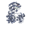
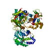
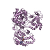
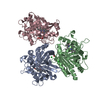
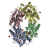
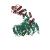
| Title | Crystal Structure of Mtb FadD10 in Complex with Dodecanoyl-AMP | ||||||
 Components Components | Long chain fatty acid CoA ligase FadD10 | ||||||
 Keywords Keywords | TRANSFERASE / open conformation / Structural Genomics / TB Structural Genomics Consortium / TBSGC | ||||||
| Function / homology | ANL, N-terminal domain / ANL, C-terminal domain / GMP Synthetase; Chain A, domain 3 / Rossmann fold / 2-Layer Sandwich / 3-Layer(aba) Sandwich / Alpha Beta / Chem-1ZZ / :  Function and homology information Function and homology information | ||||||
| Biological species |   Mycobacterium tuberculosis (bacteria) Mycobacterium tuberculosis (bacteria) | ||||||
| Method |  X-RAY DIFFRACTION / MOLECULAR REPLACEMENT / Resolution: 2.8 Å X-RAY DIFFRACTION / MOLECULAR REPLACEMENT / Resolution: 2.8 Å | ||||||
 Authors Authors | Liu, Z. / Wang, F. / Sacchettini, J.C. / TB Structural Genomics Consortium (TBSGC) | ||||||
 Citation Citation | Journal: J.Biol.Chem. / Year: 2013 Title: Structures of Mycobacterium tuberculosis FadD10 protein reveal a new type of adenylate-forming enzyme. Authors: Liu, Z. / Ioerger, T.R. / Wang, F. / Sacchettini, J.C. | ||||||
| History |
|
- Structure visualization
| Structure viewer | Molecule: MolmilJmol/JSmol |
|---|
- Downloads & links
-Download
| PDBx/mmCIF format | 4ir7.cif.gz | 108.6 KB | Display | PDBx/mmCIF format |
|---|---|---|---|---|
| PDB format | pdb4ir7.ent.gz | 81.5 KB | Display | PDB format |
| PDBx/mmJSON format | 4ir7.json.gz | Tree view | PDBx/mmJSON format | |
| Others |  Other downloads Other downloads |
-Validation report
| Summary document | 4ir7_validation.pdf.gz | 698.7 KB | Display | wwPDB validaton report |
|---|---|---|---|---|
| Full document | 4ir7_full_validation.pdf.gz | 701.5 KB | Display | |
| Data in XML | 4ir7_validation.xml.gz | 19.5 KB | Display | |
| Data in CIF | 4ir7_validation.cif.gz | 26.1 KB | Display | |
| Arichive directory | https://data.pdbj.org/pub/pdb/validation_reports/ir/4ir7ftp://data.pdbj.org/pub/pdb/validation_reports/ir/4ir7 | HTTPS FTP |
-Related structure data


-Links
 PDBj
PDBj- Assembly
| Deposited unit | 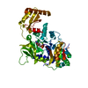
| ||||||||
|---|---|---|---|---|---|---|---|---|---|
| 1 | 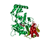
| ||||||||
| Unit cell |
|
-Components
| #1: Protein | Mass: 56727.488 Da / Num. of mol.: 1 Source method: isolated from a genetically manipulated source Source: (gene. exp.) Mycobacterium tuberculosis (bacteria) / Strain: H37Rv / Gene: Rv0099 / Plasmid: pDEST17 / Production host: |
|---|---|
| #2: Chemical | ChemComp-1ZZ /   Mass: 529.524 Da / Num. of mol.: 1 / Source method: obtained synthetically / Formula: C22H36N5O8P Mass: 529.524 Da / Num. of mol.: 1 / Source method: obtained synthetically / Formula: C22H36N5O8P |
| #3: Chemical | ChemComp-MG /   Mass: 24.305 Da / Num. of mol.: 1 / Source method: obtained synthetically / Formula: Mg Mass: 24.305 Da / Num. of mol.: 1 / Source method: obtained synthetically / Formula: Mg |
| #4: Water | ChemComp-HOH /  Mass: 18.015 Da / Num. of mol.: 32 / Source method: isolated from a natural source / Formula: H2O Mass: 18.015 Da / Num. of mol.: 32 / Source method: isolated from a natural source / Formula: H2O |
-Experimental details
-Experiment
| Experiment | Method: X-RAY DIFFRACTION / Number of used crystals: 1 |
|---|
- Sample preparation
| Crystal | Density Matthews: 4.01 Å3/Da / Density % sol: 69.36 % |
|---|---|
| Crystal grow | Temperature: 291 K / Method: vapor diffusion, hanging drop / pH: 8 Details: PEG 6000, LiSO4, pH 8.0, VAPOR DIFFUSION, HANGING DROP, temperature 291K |
-Data collection
| Diffraction | Mean temperature: 120 K | ||||||||||||||||||||||||||||||||||||||||||||||||||||||||
|---|---|---|---|---|---|---|---|---|---|---|---|---|---|---|---|---|---|---|---|---|---|---|---|---|---|---|---|---|---|---|---|---|---|---|---|---|---|---|---|---|---|---|---|---|---|---|---|---|---|---|---|---|---|---|---|---|---|
| Diffraction source | Source: ROTATING ANODE / Type: Cu FINE FOCUS / Wavelength: 1.542 Å | ||||||||||||||||||||||||||||||||||||||||||||||||||||||||
| Detector | Type: RIGAKU RAXIS / Detector: IMAGE PLATE / Date: Oct 1, 2007 | ||||||||||||||||||||||||||||||||||||||||||||||||||||||||
| Radiation | Monochromator: Ni FILTER / Protocol: SINGLE WAVELENGTH / Monochromatic (M) / Laue (L): M / Scattering type: x-ray | ||||||||||||||||||||||||||||||||||||||||||||||||||||||||
| Radiation wavelength | Wavelength: 1.542 Å / Relative weight: 1 | ||||||||||||||||||||||||||||||||||||||||||||||||||||||||
| Reflection | Resolution: 2.8→50 Å / Num. obs: 24707 / % possible obs: 99.9 % / Redundancy: 7.2 % / Rmerge(I) obs: 0.109 / Χ2: 1.603 / Net I/σ(I): 6.7 | ||||||||||||||||||||||||||||||||||||||||||||||||||||||||
| Reflection shell |
|
- Processing
| Software |
| |||||||||||||||||||||||||||||||||||||||||||||||||||||||||||||||
|---|---|---|---|---|---|---|---|---|---|---|---|---|---|---|---|---|---|---|---|---|---|---|---|---|---|---|---|---|---|---|---|---|---|---|---|---|---|---|---|---|---|---|---|---|---|---|---|---|---|---|---|---|---|---|---|---|---|---|---|---|---|---|---|---|
| Refinement | Method to determine structure: MOLECULAR REPLACEMENT / Resolution: 2.8→39.034 Å / Occupancy max: 1 / Occupancy min: 1 / FOM work R set: 0.7788 / SU ML: 0.4 / σ(F): 1.34 / Phase error: 28.43 / Stereochemistry target values: ML
| |||||||||||||||||||||||||||||||||||||||||||||||||||||||||||||||
| Solvent computation | Shrinkage radii: 0.86 Å / VDW probe radii: 1.1 Å / Solvent model: FLAT BULK SOLVENT MODEL / Bsol: 32.369 Å2 / ksol: 0.327 e/Å3 | |||||||||||||||||||||||||||||||||||||||||||||||||||||||||||||||
| Displacement parameters | Biso max: 141.69 Å2 / Biso mean: 63.6413 Å2 / Biso min: 28.3 Å2
| |||||||||||||||||||||||||||||||||||||||||||||||||||||||||||||||
| Refinement step | Cycle: LAST / Resolution: 2.8→39.034 Å
| |||||||||||||||||||||||||||||||||||||||||||||||||||||||||||||||
| Refine LS restraints |
| |||||||||||||||||||||||||||||||||||||||||||||||||||||||||||||||
| LS refinement shell | Refine-ID: X-RAY DIFFRACTION / Total num. of bins used: 8
|
