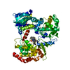
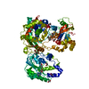
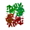
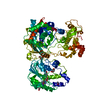
Entry Database : PDB / ID : 5jjrTitle Dengue 3 NS5 protein with compound 29 Genome polyprotein Keywords / / / Function / homology Function Domain/homology Component
/ / / / / / / / / / / / / / / / / / / / / / / / / / / / / / / / / / / / / / / / / / / / / / / / / / / / / / / / / / / / / / / / / / / / / / / / / / / / / / / / / / / / / / / / / / / / / / / / / / / / / / / / / / / / / / / / / / / / / / / / / Biological species Method / / / Resolution : 1.99 Å Authors Lescar, J. / El Sahili, A. Journal : Plos Pathog. / Year : 2016Title : Potent Allosteric Dengue Virus NS5 Polymerase Inhibitors: Mechanism of Action and Resistance ProfilingAuthors : Lim, S.P. / Noble, C.G. / Seh, C.C. / Soh, T.S. / El Sahili, A. / Chan, G.K. / Lescar, J. / Arora, R. / Benson, T. / Nilar, S. / Manjunatha, U. / Wan, K.F. / Dong, H. / Xie, X. / Shi, P.Y. / Yokokawa, F. History Deposition Apr 25, 2016 Deposition site / Processing site Revision 1.0 Jul 6, 2016 Provider / Type Revision 1.1 Aug 24, 2016 Group Revision 1.2 Nov 8, 2023 Group Data collection / Database references ... Data collection / Database references / Derived calculations / Refinement description Category chem_comp_atom / chem_comp_bond ... chem_comp_atom / chem_comp_bond / database_2 / pdbx_initial_refinement_model / pdbx_struct_conn_angle / pdbx_struct_oper_list / struct_conn Item _database_2.pdbx_DOI / _database_2.pdbx_database_accession ... _database_2.pdbx_DOI / _database_2.pdbx_database_accession / _pdbx_struct_conn_angle.ptnr1_auth_seq_id / _pdbx_struct_conn_angle.ptnr3_auth_seq_id / _pdbx_struct_conn_angle.value / _pdbx_struct_oper_list.symmetry_operation / _struct_conn.pdbx_dist_value / _struct_conn.ptnr2_auth_seq_id
Show all Show less
 Movie
Movie Controller


 Open data
 Basic information
 Components
 Keywords
 Function and homology information
 Dengue virus 3
 X-RAY DIFFRACTION /
  Authors
 Citation
 Structure visualization
 Downloads & links
 Other downloads
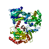


 PDBj
PDBj


 Assembly


 Mass: 65.409 Da / Num. of mol.: 2 / Source method: obtained synthetically / Formula: Zn
 Type: L-peptide linking / Mass: 384.411 Da / Num. of mol.: 1 / Source method: obtained synthetically / Formula: C14H20N6O5S
 Mass: 492.567 Da / Num. of mol.: 1 / Source method: obtained synthetically / Formula: C25H20N2O5S2
 Mass: 62.068 Da / Num. of mol.: 6 / Source method: obtained synthetically / Formula: C2H6O2
 Mass: 24.305 Da / Num. of mol.: 1 / Source method: obtained synthetically / Formula: Mg
 Mass: 106.120 Da / Num. of mol.: 3 / Source method: obtained synthetically / Formula: C4H10O3
 Sample preparation
 / Beamline: X06DA / Wavelength: 1 Å
 Processing