#191 - Nov 2015 Glutamate-gated Chloride Receptors similarity (1)
-
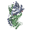
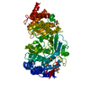
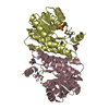
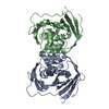
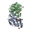
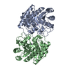
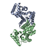
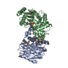
Assembly
Deposited unit
A: Asp/Glu_racemase family protein B: Asp/Glu_racemase family protein C: Asp/Glu_racemase family protein D: Asp/Glu_racemase family protein hetero molecules
Resolution: 2→29.32 Å / Cor.coef. Fo:Fc: 0.934 / Cor.coef. Fo:Fc free: 0.885 / SU B: 4.228 / SU ML: 0.121 / Cross valid method: THROUGHOUT / ESU R: 0.217 / ESU R Free: 0.187 / Stereochemistry target values: MAXIMUM LIKELIHOOD / Details: HYDROGENS HAVE BEEN ADDED IN THE RIDING POSITIONS
Rfactor
Num. reflection
% reflection
Selection details
Rfree
0.2363
2833
5.1 %
RANDOM
Rwork
0.17328
-
-
-
obs
0.17644
53262
98.1 %
-
Solvent computation
Ion probe radii: 0.8 Å / Shrinkage radii: 0.8 Å / VDW probe radii: 1.2 Å / Solvent model: MASK
 Movie
Movie Controller

 Yorodumi
 Open data
 Basic information
 Components
 Keywords
 Function and homology information
 X-RAY DIFFRACTION /
  Authors
 Citation
 Structure visualization
 Downloads & links
 Other downloads


 PDBj
PDBj

 Assembly


 Mass: 92.094 Da / Num. of mol.: 2 / Source method: obtained synthetically / Formula: C3H8O3
 Type: L-peptide linking / Mass: 147.129 Da / Num. of mol.: 3 / Source method: obtained synthetically / Formula: C5H9NO4
 Mass: 62.005 Da / Num. of mol.: 1 / Source method: obtained synthetically / Formula: NO3
 Mass: 18.015 Da / Num. of mol.: 589 / Source method: isolated from a natural source / Formula: H2O
 Sample preparation
 / Beamline: 7A (6B, 6C1) / Wavelength: 0.987 Å
 Processing