 Movie
Movie Controller

[English] 日本語
 Yorodumi
- PDB-4hzs: Crystal structure of Ack1 kinase domain with C-terminal SH3 domain -
+ Open data

- Basic information
| Entry | Database: PDB / ID: 4hzs | ||||||
|---|---|---|---|---|---|---|---|
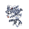
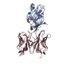
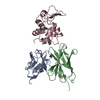
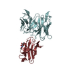
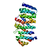
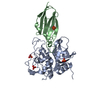
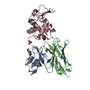
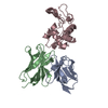
| Title | Crystal structure of Ack1 kinase domain with C-terminal SH3 domain | ||||||
 Components Components | Activated CDC42 kinase 1 | ||||||
 Keywords Keywords | TRANSFERASE / inactive state / allostery / dimerization / oligomerization / negative regulation / activation / phosphotransfer | ||||||
| Function / homology |  Function and homology information Function and homology informationregulation of clathrin-dependent endocytosis / Grb2-EGFR complex / GTPase inhibitor activity / cytoophidium / positive regulation of peptidyl-tyrosine phosphorylation / phosphorylation / clathrin-coated vesicle / Signaling by LTK / epidermal growth factor receptor binding / WW domain binding ...regulation of clathrin-dependent endocytosis / Grb2-EGFR complex / GTPase inhibitor activity / cytoophidium / positive regulation of peptidyl-tyrosine phosphorylation / phosphorylation / clathrin-coated vesicle / Signaling by LTK / epidermal growth factor receptor binding / WW domain binding / small GTPase-mediated signal transduction / protein serine/threonine/tyrosine kinase activity / clathrin-coated pit / cytoplasmic vesicle membrane / non-specific protein-tyrosine kinase / adherens junction / non-membrane spanning protein tyrosine kinase activity / endocytosis / protein tyrosine kinase activity / non-specific serine/threonine protein kinase / cell surface receptor signaling pathway / endosome / protein serine kinase activity / protein serine/threonine kinase activity / ubiquitin protein ligase binding / perinuclear region of cytoplasm / ATP binding / membrane / metal ion binding / identical protein binding / nucleus / plasma membrane / cytosol / cytoplasm Similarity search - Function | ||||||
| Biological species |  Homo sapiens (human) Homo sapiens (human) | ||||||
| Method |  X-RAY DIFFRACTION / SYNCHROTRON / MOLECULAR REPLACEMENT / Resolution: 3.23 Å X-RAY DIFFRACTION / SYNCHROTRON / MOLECULAR REPLACEMENT / Resolution: 3.23 Å | ||||||
 Authors Authors | Gajiwala, K.S. | ||||||
 Citation Citation | Journal: Plos One / Year: 2013 Title: Ack1: activation and regulation by allostery. Authors: Gajiwala, K.S. / Maegley, K. / Ferre, R. / He, Y.A. / Yu, X. | ||||||
| History |
|
- Structure visualization
| Structure viewer | Molecule: MolmilJmol/JSmol |
|---|
- Downloads & links
-Download
| PDBx/mmCIF format | 4hzs.cif.gz | 253.6 KB | Display | PDBx/mmCIF format |
|---|---|---|---|---|
| PDB format | pdb4hzs.ent.gz | 207.8 KB | Display | PDB format |
| PDBx/mmJSON format | 4hzs.json.gz | Tree view | PDBx/mmJSON format | |
| Others |  Other downloads Other downloads |
-Validation report
| Arichive directory | https://data.pdbj.org/pub/pdb/validation_reports/hz/4hzsftp://data.pdbj.org/pub/pdb/validation_reports/hz/4hzs | HTTPS FTP |
|---|
-Related structure data


-Links
 PDBj
PDBj
- Assembly
| Deposited unit | 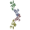
| ||||||||
|---|---|---|---|---|---|---|---|---|---|
| 1 | 
| ||||||||
| 2 | 
| ||||||||
| 3 | 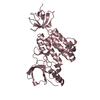
| ||||||||
| 4 | 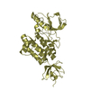
| ||||||||
| Unit cell |
|
-Components
| #1: Protein | Mass: 38861.527 Da / Num. of mol.: 4 Fragment: protein kinase and SH3 domains (UNP residues 115-453) Source method: isolated from a genetically manipulated source Source: (gene. exp.) Homo sapiens (human) / Gene: TNK2, ACK1 / Production host:   Spodoptera frugiperda (fall armyworm) Spodoptera frugiperda (fall armyworm)References: UniProt: Q07912, non-specific protein-tyrosine kinase, non-specific serine/threonine protein kinase |
|---|
-Experimental details
-Experiment
| Experiment | Method: X-RAY DIFFRACTION / Number of used crystals: 1 |
|---|
- Sample preparation
| Crystal | Density Matthews: 3.53 Å3/Da / Density % sol: 65.16 % |
|---|---|
| Crystal grow | Temperature: 286 K / Method: vapor diffusion, hanging drop / pH: 6.6 Details: 0.2 M ammonium sulfate, 0.1 M Bis-Tris, pH 6.6, 22-24% PEG3350, 10-20 mM TCEP, VAPOR DIFFUSION, HANGING DROP, temperature 286K |
-Data collection
| Diffraction | Mean temperature: 98 K |
|---|---|
| Diffraction source | Source: SYNCHROTRON / Site: APS  / Beamline: 17-ID / Wavelength: 1 Å / Beamline: 17-ID / Wavelength: 1 Å |
| Detector | Type: DECTRIS PILATUS 6M / Detector: PIXEL / Date: Mar 8, 2010 / Details: Mirrors |
| Radiation | Monochromator: Si(111) / Protocol: SINGLE WAVELENGTH / Monochromatic (M) / Laue (L): M / Scattering type: x-ray |
| Radiation wavelength | Wavelength: 1 Å / Relative weight: 1 |
| Reflection | Resolution: 3.23→73.04 Å / Num. all: 34878 / Num. obs: 34878 / % possible obs: 99.5 % / Observed criterion σ(F): 0 / Observed criterion σ(I): 0 / Redundancy: 6.5 % / Rmerge(I) obs: 0.187 / Net I/σ(I): 9.9 |
| Reflection shell | Resolution: 3.23→3.4 Å / Redundancy: 3.1 % / Rmerge(I) obs: 0.477 / Mean I/σ(I) obs: 2 / Num. unique all: 4895 / % possible all: 96.7 |
- Processing
| Software |
| ||||||||||||||||||||||||||||||||||||
|---|---|---|---|---|---|---|---|---|---|---|---|---|---|---|---|---|---|---|---|---|---|---|---|---|---|---|---|---|---|---|---|---|---|---|---|---|---|
| Refinement | Method to determine structure: MOLECULAR REPLACEMENT / Resolution: 3.23→73.04 Å / Rfactor Rfree error: 0.007 / Data cutoff high absF: 2252595.65 / Data cutoff low absF: 0 / Isotropic thermal model: RESTRAINED / Cross valid method: THROUGHOUT / σ(F): 0 / σ(I): 0 / Stereochemistry target values: Engh & Huber
| ||||||||||||||||||||||||||||||||||||
| Solvent computation | Solvent model: FLAT MODEL / Bsol: 17.8734 Å2 / ksol: 0.305827 e/Å3 | ||||||||||||||||||||||||||||||||||||
| Displacement parameters | Biso mean: 26.3 Å2
| ||||||||||||||||||||||||||||||||||||
| Refine analyze |
| ||||||||||||||||||||||||||||||||||||
| Refinement step | Cycle: LAST / Resolution: 3.23→73.04 Å
| ||||||||||||||||||||||||||||||||||||
| Refine LS restraints |
| ||||||||||||||||||||||||||||||||||||
| Refine LS restraints NCS | Weight Biso : 2 / Weight position: 300 | ||||||||||||||||||||||||||||||||||||
| LS refinement shell | Resolution: 3.23→3.43 Å / Rfactor Rfree error: 0.022 / Total num. of bins used: 6
| ||||||||||||||||||||||||||||||||||||
| Xplor file |
|
