| 登録情報 | データベース: PDB / ID: 4hwi
|
|---|
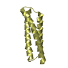
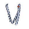
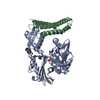
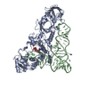
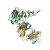
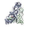
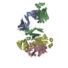
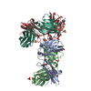
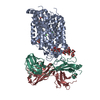
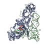
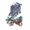
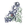
| タイトル | Crystal structure of ATBAG1 in complex with HSP70 |
|---|
 要素 要素 | - BAG family molecular chaperone regulator 1
- Heat shock cognate 71 kDa protein
|
|---|
 キーワード キーワード | CHAPERONE/APOPTOSIS / BAG domain / ubiquitin-like domain / co-chaperone / complex with molecular chaperone / CHAPERONE-APOPTOSIS complex |
|---|
| 機能・相同性 |  機能・相同性情報 機能・相同性情報
lumenal side of lysosomal membrane / regulation of protein import / negative regulation of supramolecular fiber organization / chaperone-mediated autophagy translocation complex disassembly / clathrin-sculpted gamma-aminobutyric acid transport vesicle membrane / positive regulation of ferroptosis / Lipophagy / GABA synthesis, release, reuptake and degradation / protein targeting to lysosome involved in chaperone-mediated autophagy / Respiratory syncytial virus genome transcription ...lumenal side of lysosomal membrane / regulation of protein import / negative regulation of supramolecular fiber organization / chaperone-mediated autophagy translocation complex disassembly / clathrin-sculpted gamma-aminobutyric acid transport vesicle membrane / positive regulation of ferroptosis / Lipophagy / GABA synthesis, release, reuptake and degradation / protein targeting to lysosome involved in chaperone-mediated autophagy / Respiratory syncytial virus genome transcription / adenyl-nucleotide exchange factor activity / cytoplasm protein quality control by the ubiquitin-proteasome system / clathrin coat disassembly / C3HC4-type RING finger domain binding / CHL1 interactions / regulation of protein complex stability / negative regulation of NLRP3 inflammasome complex assembly / ATP-dependent protein disaggregase activity / membrane organization / cellular response to steroid hormone stimulus / Lysosome Vesicle Biogenesis / chaperone-mediated autophagy / Golgi Associated Vesicle Biogenesis / non-chaperonin molecular chaperone ATPase / Prp19 complex / HSF1-dependent transactivation / Regulation of HSF1-mediated heat shock response / protein folding chaperone complex / response to unfolded protein / regulation of protein-containing complex assembly / Attenuation phase / ATP metabolic process / Protein methylation / heat shock protein binding / mRNA Splicing - Major Pathway / protein folding chaperone / lysosomal lumen / HSP90 chaperone cycle for steroid hormone receptors (SHR) in the presence of ligand / cellular response to starvation / spliceosomal complex / ATP-dependent protein folding chaperone / AUF1 (hnRNP D0) binds and destabilizes mRNA / regulation of protein stability / mRNA splicing, via spliceosome / Late endosomal microautophagy / PKR-mediated signaling / G protein-coupled receptor binding / Chaperone Mediated Autophagy / : / melanosome / MHC class II protein complex binding / Clathrin-mediated endocytosis / protein refolding / protein-folding chaperone binding / protein folding / Interleukin-4 and Interleukin-13 signaling / secretory granule lumen / blood microparticle / ficolin-1-rich granule lumen / protein-macromolecule adaptor activity / protein stabilization / positive regulation of cell migration / cadherin binding / ribonucleoprotein complex / receptor ligand activity / lysosomal membrane / focal adhesion / negative regulation of DNA-templated transcription / Neutrophil degranulation / ubiquitin protein ligase binding / nucleolus / enzyme binding / ATP hydrolysis activity / mitochondrion / : / RNA binding / extracellular exosome / extracellular region / nucleoplasm / ATP binding / membrane / nucleus / plasma membrane / cytoplasm / cytosol類似検索 - 分子機能 BAG domain / Molecular chaperone regulator BAG / BAG domain superfamily / BAG domain / BAG domain / BAG domain profile. / BAG domains, present in regulator of Hsp70 proteins / Defensin A-like - #30 / Defensin A-like / Heat shock hsp70 proteins family signature 2. ...BAG domain / Molecular chaperone regulator BAG / BAG domain superfamily / BAG domain / BAG domain / BAG domain profile. / BAG domains, present in regulator of Hsp70 proteins / Defensin A-like - #30 / Defensin A-like / Heat shock hsp70 proteins family signature 2. / Heat shock hsp70 proteins family signature 1. / Heat shock hsp70 proteins family signature 3. / Heat shock protein 70, conserved site / Heat shock protein 70kD, peptide-binding domain superfamily / Heat shock protein 70kD, C-terminal domain superfamily / Heat shock protein 70 family / Hsp70 protein / ATPase, substrate binding domain, subdomain 4 / Actin; Chain A, domain 4 / ATPase, nucleotide binding domain / Phosphatidylinositol 3-kinase Catalytic Subunit; Chain A, domain 1 / Methane Monooxygenase Hydroxylase; Chain G, domain 1 / ATPase, nucleotide binding domain / Ubiquitin-like (UB roll) / Nucleotidyltransferase; domain 5 / Ubiquitin domain profile. / Ubiquitin-like domain / Ubiquitin-like domain superfamily / Roll / Alpha-Beta Complex / Up-down Bundle / 2-Layer Sandwich / Mainly Alpha / Alpha Beta類似検索 - ドメイン・相同性 Heat shock cognate 71 kDa protein / BAG family molecular chaperone regulator 1類似検索 - 構成要素 |
|---|
| 生物種 |  Homo sapiens (ヒト) Homo sapiens (ヒト)
  Arabidopsis thaliana (シロイヌナズナ) Arabidopsis thaliana (シロイヌナズナ) |
|---|
| 手法 |  X線回折 / シンクロトロン / 分子置換 / 解像度: 2.273 Å X線回折 / シンクロトロン / 分子置換 / 解像度: 2.273 Å |
|---|
 データ登録者 データ登録者 | Shen, Y. / Fang, S. |
|---|
 引用 引用 | ジャーナル: Acta Crystallogr.,Sect.D / 年: 2013
タイトル: Structural insight into plant programmed cell death mediated by BAG proteins in Arabidopsis thaliana.
著者: Fang, S. / Li, L. / Cui, B. / Men, S. / Shen, Y. / Yang, X. |
|---|
| 履歴 | | 登録 | 2012年11月7日 | 登録サイト: RCSB / 処理サイト: RCSB |
|---|
| 改定 1.0 | 2013年5月29日 | Provider: repository / タイプ: Initial release |
|---|
| 改定 1.1 | 2013年6月19日 | Group: Database references |
|---|
| 改定 1.2 | 2023年9月20日 | Group: Data collection / Database references / Refinement description
カテゴリ: chem_comp_atom / chem_comp_bond ...chem_comp_atom / chem_comp_bond / database_2 / pdbx_initial_refinement_model / struct_ref_seq_dif
Item: _database_2.pdbx_DOI / _database_2.pdbx_database_accession / _struct_ref_seq_dif.details |
|---|
|
|---|
 ムービー
ムービー コントローラー


 データを開く
 基本情報
基本情報 構造の表示
構造の表示 ダウンロードとリンク
 その他のダウンロード
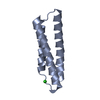


 PDBj
PDBj


 集合体
集合体

 分子量: 18.015 Da / 分子数: 114 / 由来タイプ: 天然 / 式: H2O
 試料調製
試料調製 / ビームライン: BL17U / 波長: 0.9795 / 波長: 0.9792 Å
 解析
解析