 Movie
Movie Controller


+ Open data

- Basic information
| Entry | Database: PDB / ID: 4hwd | ||||||
|---|---|---|---|---|---|---|---|
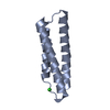
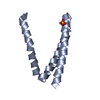
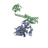
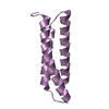
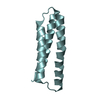
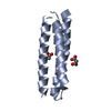
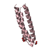
| Title | Crystal structure of ATBAG2 | ||||||
 Components Components | BAG family molecular chaperone regulator 2 | ||||||
 Keywords Keywords | APOPTOSIS / three helix bundle / co-chaperone | ||||||
| Function / homology |  Function and homology information Function and homology information | ||||||
| Biological species |  | ||||||
| Method |  X-RAY DIFFRACTION / SYNCHROTRON / MOLECULAR REPLACEMENT / Resolution: 2.312 Å X-RAY DIFFRACTION / SYNCHROTRON / MOLECULAR REPLACEMENT / Resolution: 2.312 Å | ||||||
 Authors Authors | Shen, Y. / Fang, S. | ||||||
 Citation Citation | Journal: Acta Crystallogr.,Sect.D / Year: 2013 Title: Structural insight into plant programmed cell death mediated by BAG proteins in Arabidopsis thaliana. Authors: Fang, S. / Li, L. / Cui, B. / Men, S. / Shen, Y. / Yang, X. | ||||||
| History |
|
- Structure visualization
| Structure viewer | Molecule: MolmilJmol/JSmol |
|---|
- Downloads & links
-Download
| PDBx/mmCIF format | 4hwd.cif.gz | 113.2 KB | Display | PDBx/mmCIF format |
|---|---|---|---|---|
| PDB format | pdb4hwd.ent.gz | 89.6 KB | Display | PDB format |
| PDBx/mmJSON format | 4hwd.json.gz | Tree view | PDBx/mmJSON format | |
| Others |  Other downloads Other downloads |
-Validation report
| Arichive directory | https://data.pdbj.org/pub/pdb/validation_reports/hw/4hwdftp://data.pdbj.org/pub/pdb/validation_reports/hw/4hwd | HTTPS FTP |
|---|
-Related structure data


-Links
 PDBj
PDBj- Assembly
| Deposited unit | 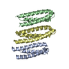
| ||||||||
|---|---|---|---|---|---|---|---|---|---|
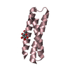
| 1 | 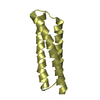
| ||||||||
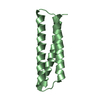
| 2 | 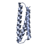
| ||||||||
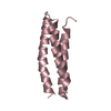
| 3 | 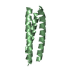
| ||||||||
| Unit cell |
|
-Components
| #1: Protein | Mass: 9920.485 Da / Num. of mol.: 3 / Fragment: BAG domain (UNP residues 129-214) Source method: isolated from a genetically manipulated source Source: (gene. exp.)  #2: Water | ChemComp-HOH / |  Mass: 18.015 Da / Num. of mol.: 182 / Source method: isolated from a natural source / Formula: H2O Mass: 18.015 Da / Num. of mol.: 182 / Source method: isolated from a natural source / Formula: H2O |
|---|
-Experimental details
-Experiment
| Experiment | Method: X-RAY DIFFRACTION / Number of used crystals: 1 |
|---|
- Sample preparation
| Crystal | Density Matthews: 2.35 Å3/Da / Density % sol: 47.65 % |
|---|---|
| Crystal grow | Temperature: 277 K / Method: vapor diffusion, sitting drop / pH: 7.5 Details: 0.2 M ammonium sulfate, 20% PEG3350, pH 7.5, VAPOR DIFFUSION, SITTING DROP, temperature 277K |
-Data collection
| Diffraction | Mean temperature: 100 K | |||||||||
|---|---|---|---|---|---|---|---|---|---|---|
| Diffraction source | Source: SYNCHROTRON / Site: SSRF  / Beamline: BL17U / Wavelength: 0.9795 / Wavelength: 0.9793 Å / Beamline: BL17U / Wavelength: 0.9795 / Wavelength: 0.9793 Å | |||||||||
| Detector | Type: RAYONIX MX-225 / Detector: CCD / Date: Feb 3, 2010 | |||||||||
| Radiation | Monochromator: double crystal Si(111) / Protocol: SINGLE WAVELENGTH / Monochromatic (M) / Laue (L): M / Scattering type: x-ray | |||||||||
| Radiation wavelength |
| |||||||||
| Reflection | Resolution: 2.312→35.711 Å / Num. all: 13096 / Num. obs: 12982 / % possible obs: 98.6 % / Observed criterion σ(F): 2 / Observed criterion σ(I): 2 | |||||||||
| Reflection shell | Resolution: 2.312→2.35 Å / % possible all: 95.3 |
- Processing
| Software |
| ||||||||||||||||||||||||||||||||||||||||||
|---|---|---|---|---|---|---|---|---|---|---|---|---|---|---|---|---|---|---|---|---|---|---|---|---|---|---|---|---|---|---|---|---|---|---|---|---|---|---|---|---|---|---|---|
| Refinement | Method to determine structure: MOLECULAR REPLACEMENT / Resolution: 2.312→35.711 Å / SU ML: 0.25 / σ(F): 0 / Phase error: 22.82 / Stereochemistry target values: ML
| ||||||||||||||||||||||||||||||||||||||||||
| Solvent computation | Shrinkage radii: 0.61 Å / VDW probe radii: 0.9 Å / Solvent model: FLAT BULK SOLVENT MODEL / Bsol: 30.304 Å2 / ksol: 0.351 e/Å3 | ||||||||||||||||||||||||||||||||||||||||||
| Displacement parameters |
| ||||||||||||||||||||||||||||||||||||||||||
| Refinement step | Cycle: LAST / Resolution: 2.312→35.711 Å
| ||||||||||||||||||||||||||||||||||||||||||
| Refine LS restraints |
| ||||||||||||||||||||||||||||||||||||||||||
| LS refinement shell |
| ||||||||||||||||||||||||||||||||||||||||||
| Refinement TLS params. | Method: refined / Origin x: -11.6405 Å / Origin y: -2.0873 Å / Origin z: 23.6578 Å
| ||||||||||||||||||||||||||||||||||||||||||
| Refinement TLS group | Selection details: all |
