 Movie
Movie Controller


+ Open data

- Basic information
| Entry | Database: PDB / ID: 4huw | ||||||
|---|---|---|---|---|---|---|---|
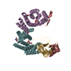
| Title | Crystal Structure of H2Db-NPM6T | ||||||
 Components Components |
| ||||||
 Keywords Keywords | IMMUNE SYSTEM / viral immunity / T cell / H2Db / influenza / viral escape | ||||||
| Function / homology |  Function and homology information Function and homology informationEndosomal/Vacuolar pathway / DAP12 interactions / Antigen Presentation: Folding, assembly and peptide loading of class I MHC / ER-Phagosome pathway / DAP12 signaling / Immunoregulatory interactions between a Lymphoid and a non-Lymphoid cell / helical viral capsid / regulation of membrane depolarization / antigen processing and presentation of peptide antigen via MHC class I / cellular defense response ...Endosomal/Vacuolar pathway / DAP12 interactions / Antigen Presentation: Folding, assembly and peptide loading of class I MHC / ER-Phagosome pathway / DAP12 signaling / Immunoregulatory interactions between a Lymphoid and a non-Lymphoid cell / helical viral capsid / regulation of membrane depolarization / antigen processing and presentation of peptide antigen via MHC class I / cellular defense response / Neutrophil degranulation / lumenal side of endoplasmic reticulum membrane / regulation of iron ion transport / cellular response to iron(III) ion / antigen processing and presentation of exogenous protein antigen via MHC class Ib, TAP-dependent / negative regulation of iron ion transport / negative regulation of forebrain neuron differentiation / regulation of erythrocyte differentiation / iron ion transport / peptide antigen assembly with MHC class I protein complex / response to molecule of bacterial origin / HFE-transferrin receptor complex / MHC class I peptide loading complex / transferrin transport / negative regulation of receptor-mediated endocytosis / cellular response to iron ion / positive regulation of T cell cytokine production / antigen processing and presentation of endogenous peptide antigen via MHC class I / MHC class I protein complex / peptide antigen assembly with MHC class II protein complex / negative regulation of neurogenesis / multicellular organismal-level iron ion homeostasis / cellular response to nicotine / MHC class II protein complex / positive regulation of receptor-mediated endocytosis / positive regulation of T cell mediated cytotoxicity / negative regulation of epithelial cell proliferation / viral penetration into host nucleus / antigen processing and presentation of exogenous peptide antigen via MHC class II / positive regulation of immune response / peptide antigen binding / phagocytic vesicle membrane / positive regulation of T cell activation / sensory perception of smell / positive regulation of cellular senescence / MHC class II protein complex binding / T cell differentiation in thymus / late endosome membrane / negative regulation of neuron projection development / host cell / antimicrobial humoral immune response mediated by antimicrobial peptide / antibacterial humoral response / cellular response to lipopolysaccharide / protein refolding / viral nucleocapsid / amyloid fibril formation / defense response to Gram-negative bacterium / protein homotetramerization / intracellular iron ion homeostasis / learning or memory / defense response to Gram-positive bacterium / immune response / ribonucleoprotein complex / external side of plasma membrane / innate immune response / lysosomal membrane / symbiont entry into host cell / host cell nucleus / structural molecule activity / Golgi apparatus / protein homodimerization activity / : / RNA binding / identical protein binding / plasma membrane / cytosol Similarity search - Function | ||||||
| Biological species |  | ||||||
| Method |  X-RAY DIFFRACTION / SYNCHROTRON / MOLECULAR REPLACEMENT / molecular replacement / Resolution: 3.16 Å X-RAY DIFFRACTION / SYNCHROTRON / MOLECULAR REPLACEMENT / molecular replacement / Resolution: 3.16 Å | ||||||
 Authors Authors | Gras, S. / Twist, K.A. / Rossjohn, J. | ||||||
 Citation Citation | Journal: Proc.Natl.Acad.Sci.USA / Year: 2013 Title: Preemptive priming readily overcomes structure-based mechanisms of virus escape. Authors: Valkenburg, S.A. / Gras, S. / Guillonneau, C. / Hatton, L.A. / Bird, N.A. / Twist, K.A. / Halim, H. / Jackson, D.C. / Purcell, A.W. / Turner, S.J. / Doherty, P.C. / Rossjohn, J. / Kedzierska, K. | ||||||
| History |
|
- Structure visualization
| Structure viewer | Molecule: MolmilJmol/JSmol |
|---|
- Downloads & links
-Download
| PDBx/mmCIF format | 4huw.cif.gz | 323.8 KB | Display | PDBx/mmCIF format |
|---|---|---|---|---|
| PDB format | pdb4huw.ent.gz | 265.2 KB | Display | PDB format |
| PDBx/mmJSON format | 4huw.json.gz | Tree view | PDBx/mmJSON format | |
| Others |  Other downloads Other downloads |
-Validation report
| Arichive directory | https://data.pdbj.org/pub/pdb/validation_reports/hu/4huwftp://data.pdbj.org/pub/pdb/validation_reports/hu/4huw | HTTPS FTP |
|---|
-Related structure data
| Related structure data |  4huuC  4huvC  4huxC  4hv8C  3cplS C: citing same article ( S: Starting model for refinement |
|---|---|
| Similar structure data |


-Links
 PDBj
PDBj


- Assembly
| Deposited unit | 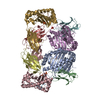
| ||||||||
|---|---|---|---|---|---|---|---|---|---|
| 1 | 
| ||||||||
| 2 | 
| ||||||||
| Unit cell |
|
-Components
| #1: Protein | Mass: 32601.303 Da / Num. of mol.: 4 Source method: isolated from a genetically manipulated source Source: (gene. exp.)  #2: Protein | Mass: 11791.545 Da / Num. of mol.: 4 Source method: isolated from a genetically manipulated source Source: (gene. exp.) #3: Protein/peptide | Mass: 996.008 Da / Num. of mol.: 4 / Source method: obtained synthetically / References: UniProt: Q5Q157*PLUS #4: Chemical | ChemComp-SO4 /   Mass: 96.063 Da / Num. of mol.: 5 / Source method: obtained synthetically / Formula: SO4 Mass: 96.063 Da / Num. of mol.: 5 / Source method: obtained synthetically / Formula: SO4#5: Water | ChemComp-HOH / |  Mass: 18.015 Da / Num. of mol.: 211 / Source method: isolated from a natural source / Formula: H2O Mass: 18.015 Da / Num. of mol.: 211 / Source method: isolated from a natural source / Formula: H2OHas protein modification | Y | |
|---|
-Experimental details
-Experiment
| Experiment | Method: X-RAY DIFFRACTION / Number of used crystals: 1 |
|---|
- Sample preparation
| Crystal | Density Matthews: 2.54 Å3/Da / Density % sol: 51.62 % |
|---|---|
| Crystal grow | Temperature: 298 K / Method: vapor diffusion, hanging drop / pH: 8.5 Details: 0.1M Tris HCl, 0.2M LiSO4, 25-30% PEG 8000, pH 8.5, vapor diffusion, hanging drop, temperature 298K |
-Data collection
| Diffraction | Mean temperature: 100 K |
|---|---|
| Diffraction source | Source: SYNCHROTRON / Site: Australian Synchrotron  / Beamline: MX2 / Wavelength: 0.956 Å / Beamline: MX2 / Wavelength: 0.956 Å |
| Detector | Type: ADSC QUANTUM 315 / Detector: CCD / Date: Apr 3, 2009 |
| Radiation | Protocol: SINGLE WAVELENGTH / Monochromatic (M) / Laue (L): M / Scattering type: x-ray |
| Radiation wavelength | Wavelength: 0.956 Å / Relative weight: 1 |
| Reflection | Resolution: 2.6→100 Å / Num. obs: 31034 / % possible obs: 94.5 % / Redundancy: 8.2 % / Biso Wilson estimate: 54.19 Å2 / Rsym value: 0.117 / Net I/σ(I): 15.5 |
-Phasing
| Phasing | Method: molecular replacement |
|---|
- Processing
| Software |
| ||||||||||||||||||||||||||||||||||||||||||||||||||||||||||||||||||||||||||||||||||||||||||||||||||||||||||||||||||
|---|---|---|---|---|---|---|---|---|---|---|---|---|---|---|---|---|---|---|---|---|---|---|---|---|---|---|---|---|---|---|---|---|---|---|---|---|---|---|---|---|---|---|---|---|---|---|---|---|---|---|---|---|---|---|---|---|---|---|---|---|---|---|---|---|---|---|---|---|---|---|---|---|---|---|---|---|---|---|---|---|---|---|---|---|---|---|---|---|---|---|---|---|---|---|---|---|---|---|---|---|---|---|---|---|---|---|---|---|---|---|---|---|---|---|---|
| Refinement | Method to determine structure: MOLECULAR REPLACEMENT Starting model: PDB entry 3cpl Resolution: 3.16→58.12 Å / Cor.coef. Fo:Fc: 0.9163 / Cor.coef. Fo:Fc free: 0.8577 / Occupancy max: 1 / Occupancy min: 0 / Cross valid method: THROUGHOUT / σ(F): 0
| ||||||||||||||||||||||||||||||||||||||||||||||||||||||||||||||||||||||||||||||||||||||||||||||||||||||||||||||||||
| Displacement parameters | Biso mean: 45.86 Å2
| ||||||||||||||||||||||||||||||||||||||||||||||||||||||||||||||||||||||||||||||||||||||||||||||||||||||||||||||||||
| Refine analyze | Luzzati coordinate error obs: 0.495 Å | ||||||||||||||||||||||||||||||||||||||||||||||||||||||||||||||||||||||||||||||||||||||||||||||||||||||||||||||||||
| Refinement step | Cycle: LAST / Resolution: 3.16→58.12 Å
| ||||||||||||||||||||||||||||||||||||||||||||||||||||||||||||||||||||||||||||||||||||||||||||||||||||||||||||||||||
| Refine LS restraints |
| ||||||||||||||||||||||||||||||||||||||||||||||||||||||||||||||||||||||||||||||||||||||||||||||||||||||||||||||||||
| LS refinement shell | Resolution: 3.16→3.26 Å / Total num. of bins used: 16
|
