 Movie
Movie Controller

[English] 日本語
 Yorodumi
- PDB-4hus: Crystal structure of streptogramin group A antibiotic acetyltrans... -
+ Open data

- Basic information
| Entry | Database: PDB / ID: 4hus | ||||||
|---|---|---|---|---|---|---|---|
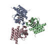
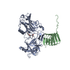
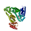
| Title | Crystal structure of streptogramin group A antibiotic acetyltransferase VatA from Staphylococcus aureus in complex with virginiamycin M1 | ||||||
 Components Components | Virginiamycin A acetyltransferase | ||||||
 Keywords Keywords | TRANSFERASE/ANTIBIOTIC / STRUCTURAL GENOMICS / ANTIBIOTIC RESISTANCE / CENTER FOR STRUCTURAL GENOMICS OF INFECTIOUS DISEASES (CSGID) / NIAID / NATIONAL INSTITUTE OF ALLERGY AND INFECTIOUS DISEASES / XENOBIOTIC ACYLTRANSFERASE (XAT) FAMILY / HEXAPEPTIDE REPEAT ACYLTRANSFERASE / streptogramin group A antibiotic acetyltransferase / STREPTOGRAMIN GROUP A ANTIBIOTICS / STREPTOGRAMIN A / VIRGINIAMYCIN M1 / DALFOPRISTIN / ACETYL COENZYME A / COENZYME A / INTRACELLULAR / TRANSFERASE-ANTIBIOTIC complex | ||||||
| Function / homology |  Function and homology information Function and homology informationacyltransferase activity / Transferases; Acyltransferases; Transferring groups other than aminoacyl groups / response to antibiotic Similarity search - Function | ||||||
| Biological species |   Staphylococcus aureus (bacteria) Staphylococcus aureus (bacteria) | ||||||
| Method |  X-RAY DIFFRACTION / SYNCHROTRON / MOLECULAR REPLACEMENT / Resolution: 2.36 Å X-RAY DIFFRACTION / SYNCHROTRON / MOLECULAR REPLACEMENT / Resolution: 2.36 Å | ||||||
 Authors Authors | Stogios, P.J. / Minasov, G. / Evdokimova, E. / Wawrzak, Z. / Yim, V. / Krishnamoorthy, M. / Di Leo, R. / Courvalin, P. / Savchenko, A. / Anderson, W.F. / Center for Structural Genomics of Infectious Diseases (CSGID) | ||||||
 Citation Citation | Journal: Antimicrob.Agents Chemother. / Year: 2014 Title: Potential for Reduction of Streptogramin A Resistance Revealed by Structural Analysis of Acetyltransferase VatA. Authors: Stogios, P.J. / Kuhn, M.L. / Evdokimova, E. / Courvalin, P. / Anderson, W.F. / Savchenko, A. | ||||||
| History |
|
- Structure visualization
| Structure viewer | Molecule: MolmilJmol/JSmol |
|---|
- Downloads & links
-Download
| PDBx/mmCIF format | 4hus.cif.gz | 286.5 KB | Display | PDBx/mmCIF format |
|---|---|---|---|---|
| PDB format | pdb4hus.ent.gz | 233 KB | Display | PDB format |
| PDBx/mmJSON format | 4hus.json.gz | Tree view | PDBx/mmJSON format | |
| Others |  Other downloads Other downloads |
-Validation report
| Arichive directory | https://data.pdbj.org/pub/pdb/validation_reports/hu/4husftp://data.pdbj.org/pub/pdb/validation_reports/hu/4hus | HTTPS FTP |
|---|
-Related structure data
| Related structure data |  4hurC  4myoC  4e8l C: citing same article ( S: Starting model for refinement |
|---|---|
| Similar structure data | |
| Other databases |


-Links
 PDBj
PDBj


- Assembly
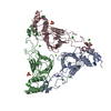
| Deposited unit | 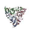
| ||||||||
|---|---|---|---|---|---|---|---|---|---|
| 1 |
| ||||||||
| Unit cell |
| ||||||||
| Components on special symmetry positions |
|
-Components
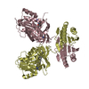
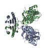
-Protein , 1 types, 3 molecules ABC
| #1: Protein | Mass: 25017.736 Da / Num. of mol.: 3 Source method: isolated from a genetically manipulated source Source: (gene. exp.) Staphylococcus aureus (bacteria) / Strain: BM3093 / Gene: vat / Plasmid: P15TV LIC / Production host: References: UniProt: P26839, Transferases; Acyltransferases; Transferring groups other than aminoacyl groups |
|---|
-Non-polymers , 8 types, 548 molecules 


| #2: Chemical | ChemComp-SO4 /  Mass: 96.063 Da / Num. of mol.: 8 / Source method: obtained synthetically / Formula: SO4 Mass: 96.063 Da / Num. of mol.: 8 / Source method: obtained synthetically / Formula: SO4#3: Chemical | ChemComp-NA / |  Mass: 22.990 Da / Num. of mol.: 1 / Source method: obtained synthetically / Formula: Na Mass: 22.990 Da / Num. of mol.: 1 / Source method: obtained synthetically / Formula: Na#4: Chemical | ChemComp-CL /  Mass: 35.453 Da / Num. of mol.: 9 / Source method: obtained synthetically / Formula: Cl Mass: 35.453 Da / Num. of mol.: 9 / Source method: obtained synthetically / Formula: Cl#5: Chemical | ChemComp-EDO /  Mass: 62.068 Da / Num. of mol.: 15 / Source method: obtained synthetically / Formula: C2H6O2 Mass: 62.068 Da / Num. of mol.: 15 / Source method: obtained synthetically / Formula: C2H6O2#6: Chemical | ChemComp-PEG /  Mass: 106.120 Da / Num. of mol.: 4 / Source method: obtained synthetically / Formula: C4H10O3 Mass: 106.120 Da / Num. of mol.: 4 / Source method: obtained synthetically / Formula: C4H10O3#7: Chemical | ChemComp-VIR / |  Mass: 525.593 Da / Num. of mol.: 1 / Source method: obtained synthetically / Formula: C28H35N3O7 / Comment: antibiotic*YM Mass: 525.593 Da / Num. of mol.: 1 / Source method: obtained synthetically / Formula: C28H35N3O7 / Comment: antibiotic*YM#8: Chemical |  Mass: 150.173 Da / Num. of mol.: 2 / Source method: obtained synthetically / Formula: C6H14O4 Mass: 150.173 Da / Num. of mol.: 2 / Source method: obtained synthetically / Formula: C6H14O4#9: Water | ChemComp-HOH / | Mass: 18.015 Da / Num. of mol.: 508 / Source method: isolated from a natural source / Formula: H2O |
|---|
-Experimental details
-Experiment
| Experiment | Method: X-RAY DIFFRACTION / Number of used crystals: 1 |
|---|
- Sample preparation
| Crystal | Density Matthews: 2.83 Å3/Da / Density % sol: 56.53 % |
|---|---|
| Crystal grow | Temperature: 298 K / Method: vapor diffusion, sitting drop / pH: 7.5 Details: 2 M ammonium sulphate, 0.1 M HEPES pH 7.5, 2% PEG 400, virginiamycin M1, VAPOR DIFFUSION, SITTING DROP, temperature 298K |
-Data collection
| Diffraction | Mean temperature: 100 K |
|---|---|
| Diffraction source | Source: SYNCHROTRON / Site: APS  / Beamline: 21-ID-F / Wavelength: 0.97872 Å / Beamline: 21-ID-F / Wavelength: 0.97872 Å |
| Detector | Type: MARMOSAIC 225 mm CCD / Detector: CCD / Date: Jul 27, 2012 |
| Radiation | Monochromator: C(111) / Protocol: SINGLE WAVELENGTH / Monochromatic (M) / Laue (L): M / Scattering type: x-ray |
| Radiation wavelength | Wavelength: 0.97872 Å / Relative weight: 1 |
| Reflection | Resolution: 2.3→30 Å / Num. obs: 37898 / % possible obs: 99.1 % / Observed criterion σ(F): 0 / Observed criterion σ(I): -2 / Redundancy: 6 % / Rsym value: 0.079 / Net I/σ(I): 25.68 |
| Reflection shell | Resolution: 2.3→2.34 Å / Redundancy: 5.8 % / Mean I/σ(I) obs: 2.64 / Rsym value: 0.576 / % possible all: 100 |
- Processing
| Software |
| ||||||||||||||||||||||||||||||||||||||||||||||||||||||||||||||||||||||||||||||||||||||||||||||||||||||||||||||||||||||||||||||||||||||||||||||||||||||||||||||||||||||||||||||||||||||||||||||||||||||||||||||||||||||||||||||||||||||||||||||||||||||||||
|---|---|---|---|---|---|---|---|---|---|---|---|---|---|---|---|---|---|---|---|---|---|---|---|---|---|---|---|---|---|---|---|---|---|---|---|---|---|---|---|---|---|---|---|---|---|---|---|---|---|---|---|---|---|---|---|---|---|---|---|---|---|---|---|---|---|---|---|---|---|---|---|---|---|---|---|---|---|---|---|---|---|---|---|---|---|---|---|---|---|---|---|---|---|---|---|---|---|---|---|---|---|---|---|---|---|---|---|---|---|---|---|---|---|---|---|---|---|---|---|---|---|---|---|---|---|---|---|---|---|---|---|---|---|---|---|---|---|---|---|---|---|---|---|---|---|---|---|---|---|---|---|---|---|---|---|---|---|---|---|---|---|---|---|---|---|---|---|---|---|---|---|---|---|---|---|---|---|---|---|---|---|---|---|---|---|---|---|---|---|---|---|---|---|---|---|---|---|---|---|---|---|---|---|---|---|---|---|---|---|---|---|---|---|---|---|---|---|---|---|---|---|---|---|---|---|---|---|---|---|---|---|---|---|---|---|---|---|---|---|---|---|---|---|---|---|---|---|---|---|---|---|
| Refinement | Method to determine structure: MOLECULAR REPLACEMENT Starting model: 4E8L 4e8l Resolution: 2.36→29.389 Å / SU ML: 0.2 / σ(F): 1.34 / Phase error: 24.63 / Stereochemistry target values: ML
| ||||||||||||||||||||||||||||||||||||||||||||||||||||||||||||||||||||||||||||||||||||||||||||||||||||||||||||||||||||||||||||||||||||||||||||||||||||||||||||||||||||||||||||||||||||||||||||||||||||||||||||||||||||||||||||||||||||||||||||||||||||||||||
| Solvent computation | Shrinkage radii: 0.9 Å / VDW probe radii: 1.11 Å / Solvent model: FLAT BULK SOLVENT MODEL | ||||||||||||||||||||||||||||||||||||||||||||||||||||||||||||||||||||||||||||||||||||||||||||||||||||||||||||||||||||||||||||||||||||||||||||||||||||||||||||||||||||||||||||||||||||||||||||||||||||||||||||||||||||||||||||||||||||||||||||||||||||||||||
| Refinement step | Cycle: LAST / Resolution: 2.36→29.389 Å
| ||||||||||||||||||||||||||||||||||||||||||||||||||||||||||||||||||||||||||||||||||||||||||||||||||||||||||||||||||||||||||||||||||||||||||||||||||||||||||||||||||||||||||||||||||||||||||||||||||||||||||||||||||||||||||||||||||||||||||||||||||||||||||
| Refine LS restraints |
| ||||||||||||||||||||||||||||||||||||||||||||||||||||||||||||||||||||||||||||||||||||||||||||||||||||||||||||||||||||||||||||||||||||||||||||||||||||||||||||||||||||||||||||||||||||||||||||||||||||||||||||||||||||||||||||||||||||||||||||||||||||||||||
| LS refinement shell |
| ||||||||||||||||||||||||||||||||||||||||||||||||||||||||||||||||||||||||||||||||||||||||||||||||||||||||||||||||||||||||||||||||||||||||||||||||||||||||||||||||||||||||||||||||||||||||||||||||||||||||||||||||||||||||||||||||||||||||||||||||||||||||||
| Refinement TLS params. | Method: refined / Refine-ID: X-RAY DIFFRACTION
| ||||||||||||||||||||||||||||||||||||||||||||||||||||||||||||||||||||||||||||||||||||||||||||||||||||||||||||||||||||||||||||||||||||||||||||||||||||||||||||||||||||||||||||||||||||||||||||||||||||||||||||||||||||||||||||||||||||||||||||||||||||||||||
| Refinement TLS group |
|
