 Movie
Movie Controller

[English] 日本語
 Yorodumi
- PDB-4hn1: Crystal Structure of H60N/Y130F double mutant of ChmJ, a 3'-monoe... -
+ Open data

- Basic information
| Entry | Database: PDB / ID: 4hn1 | ||||||
|---|---|---|---|---|---|---|---|
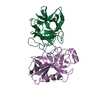
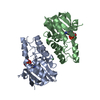
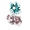
| Title | Crystal Structure of H60N/Y130F double mutant of ChmJ, a 3'-monoepimerase from Streptomyces bikiniensis in complex with dTDP | ||||||
 Components Components | Putative 3-epimerase in D-allose pathway | ||||||
 Keywords Keywords | UNKNOWN FUNCTION / 3'-monoepimerase / natural product / deoxysugar / chalcomycin / dTDP-mycinose / Cupin fold / nucleotide-linked sugar / Epimerization | ||||||
| Function / homology |  Function and homology information Function and homology informationdTDP-4-dehydro-6-deoxy-D-glucose 3-epimerase / dTDP-4-dehydrorhamnose 3,5-epimerase activity / dTDP-rhamnose biosynthetic process / polysaccharide biosynthetic process / antibiotic biosynthetic process / cytosol Similarity search - Function | ||||||
| Biological species |  Streptomyces bikiniensis (bacteria) Streptomyces bikiniensis (bacteria) | ||||||
| Method |  X-RAY DIFFRACTION / SYNCHROTRON / MOLECULAR REPLACEMENT / Resolution: 1.6 Å X-RAY DIFFRACTION / SYNCHROTRON / MOLECULAR REPLACEMENT / Resolution: 1.6 Å | ||||||
 Authors Authors | Holden, H.M. / Kubiak, R.L. | ||||||
 Citation Citation | Journal: Biochemistry / Year: 2012 Title: Structural and Functional Studies on a 3'-Epimerase Involved in the Biosynthesis of dTDP-6-deoxy-d-allose. Authors: Kubiak, R.L. / Phillips, R.K. / Zmudka, M.W. / Ahn, M.R. / Maka, E.M. / Pyeatt, G.L. / Roggensack, S.J. / Holden, H.M. | ||||||
| History |
|
- Structure visualization
| Structure viewer | Molecule: MolmilJmol/JSmol |
|---|
- Downloads & links
-Download
| PDBx/mmCIF format | 4hn1.cif.gz | 180.7 KB | Display | PDBx/mmCIF format |
|---|---|---|---|---|
| PDB format | pdb4hn1.ent.gz | 144.5 KB | Display | PDB format |
| PDBx/mmJSON format | 4hn1.json.gz | Tree view | PDBx/mmJSON format | |
| Others |  Other downloads Other downloads |
-Validation report
| Arichive directory | https://data.pdbj.org/pub/pdb/validation_reports/hn/4hn1ftp://data.pdbj.org/pub/pdb/validation_reports/hn/4hn1 | HTTPS FTP |
|---|
-Related structure data
| Related structure data |  4hmzSC  4hn0C S: Starting model for refinement C: citing same article ( |
|---|---|
| Similar structure data |


-Links
 PDBj
PDBj- Assembly
| Deposited unit | 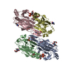
| ||||||||
|---|---|---|---|---|---|---|---|---|---|
| 1 | 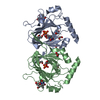
| ||||||||
| 2 | 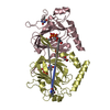
| ||||||||
| Unit cell |
|
-Components
-Protein , 1 types, 4 molecules ABCD
| #1: Protein | Mass: 22323.840 Da / Num. of mol.: 4 / Mutation: H60N, Y130F Source method: isolated from a genetically manipulated source Source: (gene. exp.) Streptomyces bikiniensis (bacteria) / Gene: chmJ / Plasmid: pET-31b(+) / Production host: |
|---|
-Non-polymers , 5 types, 438 molecules 


| #2: Chemical | ChemComp-EDO /  Mass: 62.068 Da / Num. of mol.: 11 / Source method: obtained synthetically / Formula: C2H6O2 Mass: 62.068 Da / Num. of mol.: 11 / Source method: obtained synthetically / Formula: C2H6O2#3: Chemical | ChemComp-TYD /  Mass: 402.188 Da / Num. of mol.: 4 / Source method: obtained synthetically / Formula: C10H16N2O11P2 Mass: 402.188 Da / Num. of mol.: 4 / Source method: obtained synthetically / Formula: C10H16N2O11P2#4: Chemical |  Type: DNA OH 5 prime terminus / Mass: 242.229 Da / Num. of mol.: 2 / Source method: obtained synthetically / Formula: C10H14N2O5 Type: DNA OH 5 prime terminus / Mass: 242.229 Da / Num. of mol.: 2 / Source method: obtained synthetically / Formula: C10H14N2O5#5: Chemical |  Mass: 126.113 Da / Num. of mol.: 2 / Source method: obtained synthetically / Formula: C5H6N2O2 Mass: 126.113 Da / Num. of mol.: 2 / Source method: obtained synthetically / Formula: C5H6N2O2#6: Water | ChemComp-HOH / | Mass: 18.015 Da / Num. of mol.: 419 / Source method: isolated from a natural source / Formula: H2O |
|---|
-Experimental details
-Experiment
| Experiment | Method: X-RAY DIFFRACTION / Number of used crystals: 1 |
|---|
- Sample preparation
| Crystal | Density Matthews: 3.24 Å3/Da / Density % sol: 62.05 % |
|---|---|
| Crystal grow | Temperature: 298 K / Method: vapor diffusion, hanging drop / pH: 6.5 Details: 20% PEG 3400, 2% 3-methyl-1,5-pentanediol, 100 mM MOPS, pH 6.5, VAPOR DIFFUSION, HANGING DROP, temperature 298K |
-Data collection
| Diffraction | Mean temperature: 100 K |
|---|---|
| Diffraction source | Source: SYNCHROTRON / Site: APS  / Beamline: 19-BM / Wavelength: 0.979 Å / Beamline: 19-BM / Wavelength: 0.979 Å |
| Detector | Type: ADSC QUANTUM 210r / Detector: CCD / Date: Aug 4, 2012 |
| Radiation | Monochromator: double crystal monochromator Si(111) / Protocol: SINGLE WAVELENGTH / Monochromatic (M) / Laue (L): M / Scattering type: x-ray |
| Radiation wavelength | Wavelength: 0.979 Å / Relative weight: 1 |
| Reflection | Resolution: 1.6→99.71 Å / Num. all: 143838 / Num. obs: 143838 / % possible obs: 96.1 % / Redundancy: 4.9 % |
| Reflection shell | Resolution: 1.6→1.66 Å / Redundancy: 2.8 % / Mean I/σ(I) obs: 2.07 / Num. unique all: 13940 / Rsym value: 0.39 / % possible all: 93.6 |
- Processing
| Software |
| |||||||||||||||||||||||||||||||||||||||||||||||||||||||||||||||||
|---|---|---|---|---|---|---|---|---|---|---|---|---|---|---|---|---|---|---|---|---|---|---|---|---|---|---|---|---|---|---|---|---|---|---|---|---|---|---|---|---|---|---|---|---|---|---|---|---|---|---|---|---|---|---|---|---|---|---|---|---|---|---|---|---|---|---|
| Refinement | Method to determine structure: MOLECULAR REPLACEMENT Starting model: PDB entry 4HMZ Resolution: 1.6→30 Å / Cor.coef. Fo:Fc: 0.958 / Cor.coef. Fo:Fc free: 0.938 / SU B: 2.074 / SU ML: 0.071 / Cross valid method: THROUGHOUT / ESU R: 0.088 / ESU R Free: 0.094 / Stereochemistry target values: MAXIMUM LIKELIHOOD / Details: HYDROGENS HAVE BEEN ADDED IN THE RIDING POSITIONS
| |||||||||||||||||||||||||||||||||||||||||||||||||||||||||||||||||
| Solvent computation | Ion probe radii: 0.8 Å / Shrinkage radii: 0.8 Å / VDW probe radii: 1.4 Å / Solvent model: MASK | |||||||||||||||||||||||||||||||||||||||||||||||||||||||||||||||||
| Displacement parameters | Biso mean: 29.798 Å2
| |||||||||||||||||||||||||||||||||||||||||||||||||||||||||||||||||
| Refinement step | Cycle: LAST / Resolution: 1.6→30 Å
| |||||||||||||||||||||||||||||||||||||||||||||||||||||||||||||||||
| Refine LS restraints |
| |||||||||||||||||||||||||||||||||||||||||||||||||||||||||||||||||
| LS refinement shell | Resolution: 1.6→1.641 Å / Total num. of bins used: 20
|
